XSP-8C Series Biological Microscope
| Origin | Shanghai, China |
|---|---|
| Manufacturer Type | Authorized Distributor |
| Origin Category | Domestic (PRC) |
| Model | XSP-8C Series |
| Pricing | Available Upon Request |
| Eyepieces | WF10×/18 mm, WF16×/11 mm |
| Objectives | Plan Achromatic 4× (NA 0.10, WD 17.5 mm), 10× (NA 0.25, WD 7.31 mm), 40× (NA 0.65, WD 0.63 mm), 100× Oil (NA 1.25, WD 0.18 mm) |
| Total Optical Magnification | 40×–1600× |
| System Reference Magnification | Up to 2600× (with eyepiece & objective combination) |
| Focusing Mechanism | Coaxial coarse/fine focus, 15 mm travel, fine adjustment graduation 2 µm |
| Mechanical Tube Length | 160 mm |
| Condenser | NA 1.25 Abbe condenser with iris diaphragm |
| Stage | Mechanical stage 150 × 135 mm, X–Y travel 75 × 50 mm, vernier scale 0.1 mm |
| Illumination | 6 V / 20 W halogen lamp, 110–220 V AC input, continuously variable brightness control |
| Filter Set | Blue, green, yellow interference filters |
| Anti-fungal Treatment | Integrated optical component anti-mold coating |
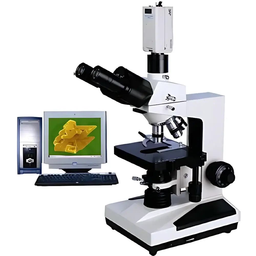
| Imaging Configurations | XSP-8CE (CCD-based computer system), XSP-8CZ (digital camera–coupled system) |
| Display-Based Max Magnification | 2800× (17″ monitor, XSP-8CE), 2400× (XSP-8CZ with standard DSLR) |
Overview
The XSP-8C Series Biological Microscope is a precision-engineered upright transmitted-light microscope designed for routine and advanced life science applications in academic laboratories, clinical diagnostics, pharmaceutical research, and industrial quality control. Built on a robust 160 mm mechanical tube length platform, it adheres to classical finite-conjugate optical design principles and supports standardized plan achromatic objectives compliant with international microscopy conventions. Its optical architecture enables high-fidelity brightfield observation across a magnification range of 40× to 1600×, extendable to 2600× under optimal eyepiece–objective pairing. The instrument employs Köhler illumination methodology via an NA 1.25 Abbe condenser and a continuously adjustable 6 V / 20 W halogen light source—ensuring uniform, glare-free illumination critical for reproducible morphological assessment and quantitative image capture. All optical components—including prisms, lenses, and eyepiece field stops—are treated with proprietary anti-fungal coatings to ensure long-term stability in humid laboratory environments common to tropical or subtropical regions.
Key Features
- Coaxial coarse and fine focusing mechanism with 15 mm vertical travel and 2 µm fine-focus graduation for precise Z-axis positioning and repeatable focal plane registration.
- Dual-widefield eyepieces (WF10×/18 mm and WF16×/11 mm) offering ergonomic viewing angles and extended eye relief; binocular head rotates 360° for collaborative observation or space-constrained benchtop integration.
- Four-position revolving nosepiece accommodating plan achromatic objectives (4×, 10×, 40×, and oil-immersion 100×), each optimized for chromatic and spherical aberration correction at specified working distances.
- Large mechanical stage (150 × 135 mm) with calibrated X–Y translation (75 × 50 mm range) and 0.1 mm vernier readout—enabling systematic scanning of histological sections, microbial cultures, or tissue arrays.
- Integrated filter drawer supporting blue, green, and yellow interference filters for contrast enhancement in unstained or lightly stained specimens without requiring external accessories.
- Universal power supply (110–220 V AC) with dimmable halogen illumination—compatible with global electrical infrastructure and suitable for GLP-compliant documentation workflows where consistent lighting intensity must be logged and verified.
Sample Compatibility & Compliance
The XSP-8C Series accommodates standard 1 mm-thick glass microscope slides and 22 × 22 mm coverslips, supporting both fixed and live specimens—including hematology smears, bacterial Gram preparations, plant epidermal peels, and cultured mammalian monolayers. Optional contrast-enhancement modules—including phase contrast, darkfield, and polarizing attachments—can be retrofitted without optical realignment, enabling method flexibility per ISO 10993-12 (biocompatibility evaluation) or CLSI EP17-A2 (microscopy-based cell enumeration). While not certified for IVD use under EU IVDR or FDA 510(k), the system meets mechanical and electrical safety requirements per IEC 61010-1:2010 for laboratory equipment. Its modular construction facilitates audit-ready configuration management in GMP-aligned QC labs performing raw material inspection or stability testing per USP or .
Software & Data Management
The XSP-8CE (computer-coupled) and XSP-8CZ (camera-coupled) configurations support vendor-agnostic digital imaging workflows. The XSP-8CE variant integrates a CCD sensor with USB 2.0 A/D converter and includes basic acquisition software compatible with Windows 10/11; metadata tagging (date/time, objective used, exposure settings) is embedded into TIFF and BMP exports. For regulated environments, third-party image analysis platforms—such as ImageJ/Fiji (NIH), HALO (Indica Labs), or Olympus cellSens—can be validated per 21 CFR Part 11 when deployed with appropriate electronic signature and audit trail protocols. All digital outputs retain native pixel resolution without interpolation, preserving fidelity required for peer-reviewed publication or regulatory submission.
Applications
- Academic instruction: Morphological study of mitotic stages, protozoan motility, fungal hyphae, and vascular anatomy in undergraduate biology curricula.
- Clinical pathology: Routine examination of peripheral blood films, Pap smears, and biopsy touch preps in hospital laboratories operating under CAP-accredited standards.
- Pharmaceutical microbiology: Enumeration of environmental isolates per USP , detection of particulate contaminants in parenteral solutions, and verification of sterility test membrane filtration results.
- Agricultural biotechnology: Pollen viability assessment, seed coat ultrastructure analysis, and nematode identification in soil extracts.
- Industrial QA: Inspection of filter integrity, fiber morphology in textile composites, and crystallization behavior in active pharmaceutical ingredient (API) slurries.
FAQ
Is the XSP-8C Series compatible with oil immersion objectives?
Yes—the 100× plan achromatic oil objective (NA 1.25, WD 0.18 mm) is factory-aligned and included in the standard configuration. Immersion oil with refractive index 1.515 is recommended.
Can this microscope be used for fluorescence observation?
No—fluorescence capability requires epi-illumination, dichroic mirrors, and excitation/emission filter sets, none of which are supported by the XSP-8C’s transmitted-light-only optical path.
What is the maximum digital magnification achievable with the XSP-8CE system?
When displayed on a 17″ monitor at native resolution (1280 × 1024), the system achieves up to 2800× reference magnification—calculated using sensor pixel size, monitor pixel pitch, and optical magnification factors per ISO 8578:2017.
Does the anti-mold treatment cover all optical surfaces?
Yes—the proprietary coating is applied to all air-to-glass interfaces within the optical train, including eyepiece lenses, prism assemblies, and objective rear elements, and remains effective for ≥5 years under typical lab humidity conditions (40–60% RH).
Is technical documentation available in English for regulatory submission purposes?
Full English-language user manuals, calibration certificates (traceable to NIM, China), and CE Declaration of Conformity (for distributor-supplied units meeting EN 61010-1) are provided upon order confirmation.
Related Products


