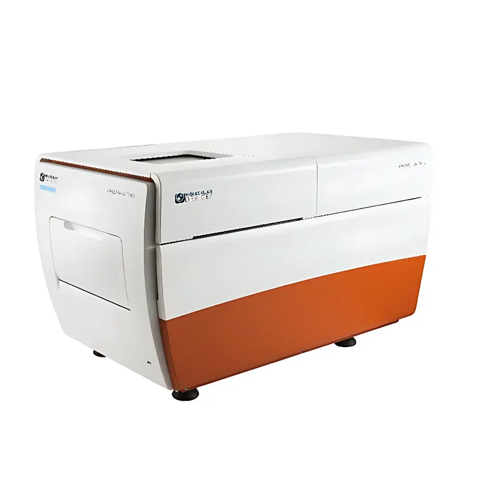
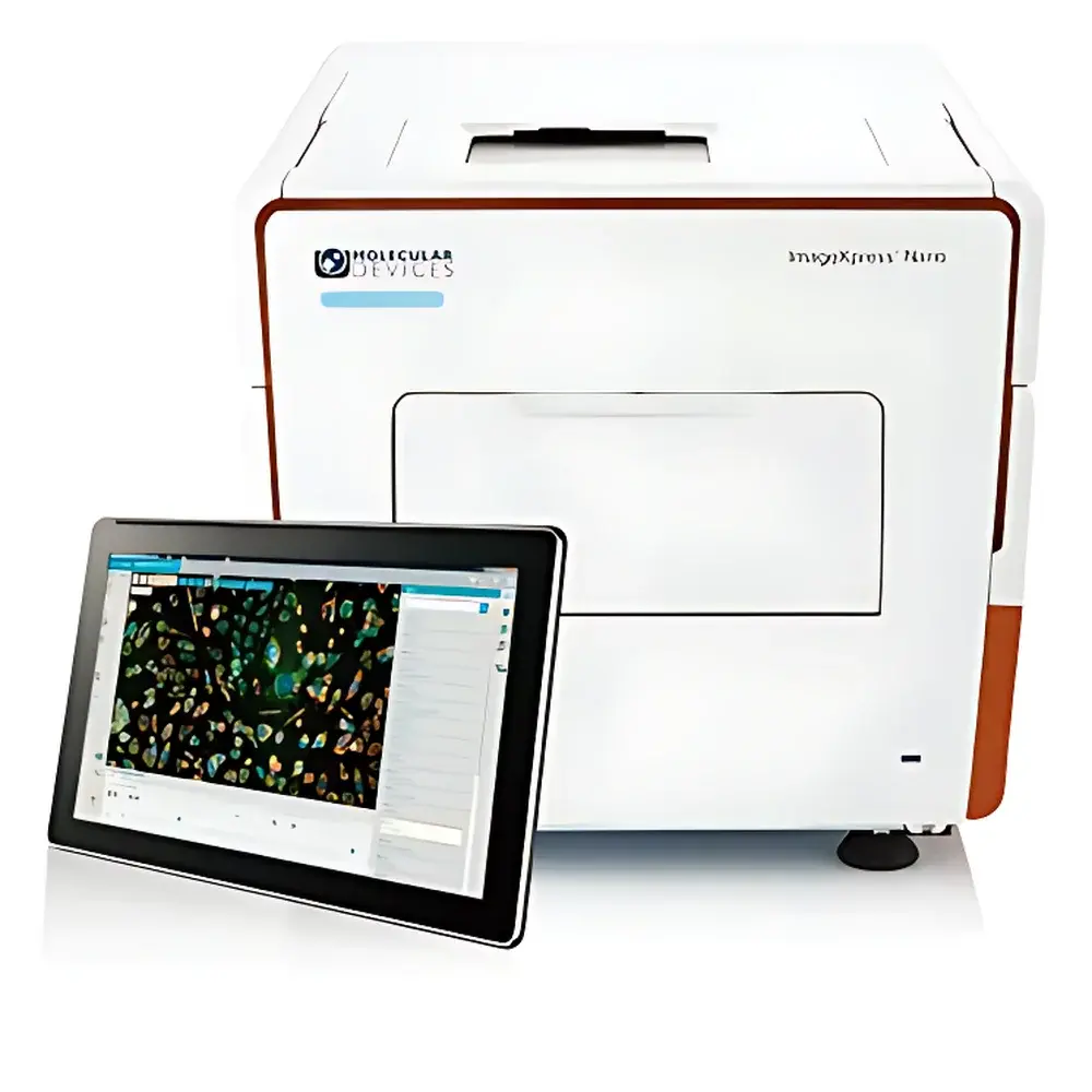
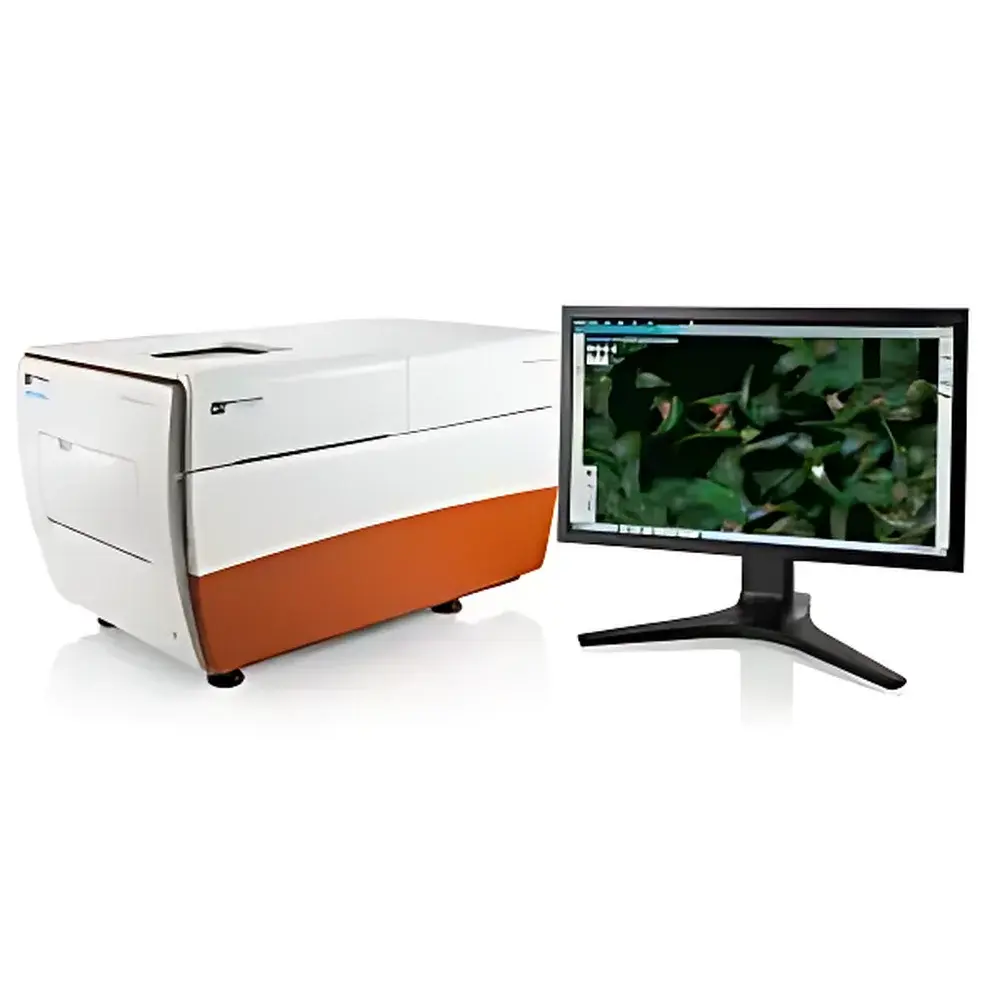
Molecular Devices ImageXpress Nano Automated High-Content Imaging System
| Brand | Molecular Devices |
|---|---|
| Origin | USA |
| Manufacturer Type | Original Equipment Manufacturer (OEM) |
| Origin Category | Imported |
| Model | ImageXpress Nano |
| Pricing | Available upon request |
Overview
The Molecular Devices ImageXpress Nano Automated High-Content Imaging System is a benchtop, fully automated fluorescence and brightfield microscope platform engineered for quantitative cellular phenotyping at microplate and slide scale. It employs widefield epifluorescence and transmitted-light imaging principles—optimized via high-quantum-efficiency sCMOS sensors, precision motorized stages, and solid-state LED illumination—to deliver reproducible, high-fidelity image acquisition across 6–384-well plates and standard glass slides. Designed for laboratories transitioning from manual microscopy to scalable, standardized cell-based assays, the system bridges the gap between research-grade imaging and operational robustness required in preclinical screening, toxicology, and functional genomics workflows. Its architecture supports label-free brightfield analysis (e.g., phase contrast-equivalent segmentation), multiplexed fluorescence detection (with user-selectable filter cubes and objectives), and optional environmental control (temperature, CO2, humidity) for live-cell kinetics.
Key Features
- Integrated solid-state LED light engine with stable spectral output and >20,000-hour lifetime—minimizing phototoxicity and intensity drift during time-lapse experiments.
- Motorized, high-precision XYZ stage with sub-micron repeatability for consistent multi-position acquisition across wells or slide regions.
- Modular optical path supporting interchangeable objectives (1.25× to 60×), emission filter sets, and dual-channel simultaneous imaging capability.
- CellReporterXpress™ software with embedded 25 pre-validated analysis modules—including brightfield object recognition (small round cells, large adherent cells, beads), nuclear/cytoplasmic segmentation, and intensity-based quantification of fluorescent reporters.
- Touch-optimized interface compatible with Windows tablets and desktops; full remote operation via Chrome or Safari browsers without client-side installation.
- CellMagnify™ technology enabling seamless zoom-from-plate to single-cell resolution with synchronized panning—supporting side-by-side comparative analysis of two wells or fields-of-view.
- Region-of-interest (ROI) acquisition mode allowing targeted imaging of up to 999 unique locations per well or slide—ideal for heterogeneous samples or spatially resolved assays.
Sample Compatibility & Compliance
The ImageXpress Nano accommodates standard ANSI/SLAS-compliant microplates (6–384-well), chambered coverslips, and 1″ × 3″ glass slides. It supports fixed and live-cell assays—including immunofluorescence, GFP/RFP reporter systems, viability dyes (e.g., Calcein AM/Propidium Iodide), and label-free morphological profiling. Environmental control options meet ASTM E1973-22 requirements for CO2 regulation and ISO 15197:2013 humidity stability thresholds. Data integrity complies with FDA 21 CFR Part 11 through audit-trail-enabled user authentication, electronic signatures, and immutable acquisition metadata logging. The system is validated for GLP/GMP-aligned workflows and integrates with LIMS and ELN platforms via standard DICOM and TIFF export protocols.
Software & Data Management
CellReporterXpress™ provides a unified workflow environment—from acquisition protocol definition to pixel-level analysis and statistical reporting. All analysis pipelines are scriptable using Python-based APIs and support batch reprocessing across historical datasets. Distributed image analysis leverages local multicore processing and optional cloud-offload for accelerated throughput—enabling >100 plates/day in typical assay configurations. Raw images are stored in OME-TIFF format with embedded acquisition parameters (exposure time, gain, Z-stack intervals, objective ID). Export options include CSV (per-object measurements), PDF reports with QC metrics (focus score, signal-to-noise ratio), and interactive HTML viewers for collaborative review. Audit trails record operator actions, parameter changes, and software version history—fully traceable for regulatory submissions.
Applications
- High-content cytotoxicity screening (e.g., mitochondrial membrane potential loss, lysosomal permeabilization, caspase activation).
- Neurite outgrowth and synapse quantification in iPSC-derived neuronal models.
- Cell cycle profiling via DNA content and phospho-histone H3 co-staining.
- Organoid size, morphology, and lumen formation analysis in 3D culture systems.
- CRISPR screen hit validation using multiplexed marker expression and subcellular localization metrics.
- Label-free confluence monitoring and wound-healing assay kinetics under controlled environmental conditions.
FAQ
Does the ImageXpress Nano support Z-stack acquisition and deconvolution?
Yes—it acquires Z-series with programmable step size (0.1–20 µm) and integrates GPU-accelerated widefield deconvolution using constrained iterative algorithms.
Can I import custom analysis algorithms developed in MATLAB or Python?
Yes—CellReporterXpress supports third-party plugin integration via its open API framework and allows execution of external scripts as analysis modules.
Is environmental control available as a field-upgradeable option?
Yes—CO2/humidity/temperature modules can be installed post-purchase with factory calibration and IQ/OQ documentation.
What file formats are supported for data export and long-term archiving?
OME-TIFF (primary), TIFF, PNG, CSV, PDF, and JSON metadata files—all compliant with NIH BioImage Archive submission standards.
How does the system ensure focus stability across multi-day time-lapse experiments?
It uses real-time hardware autofocus based on contrast gradient maximization at each Z-plane, recalibrated before every field acquisition and logged in audit trail records.