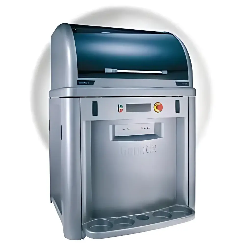
Molecular Devices ClonePix 2 High-Throughput Monoclonal Cell Screening System
| Brand | Molecular Devices |
|---|---|
| Origin | Shanghai, China |
| Manufacturer Type | Original Equipment Manufacturer (OEM) |
| Product Origin | Domestic (China) |
| Model | ClonePix 2 |
| Pricing | Available upon Request |
Overview
The Molecular Devices ClonePix 2 High-Throughput Monoclonal Cell Screening System is an automated imaging and picking platform engineered for precision monoclonal antibody (mAb) and recombinant protein-producing cell line development. It employs dual-modality digital imaging—white-light and fluorescence—to quantify clone morphology (size, circularity, proximity) and target protein expression levels directly within semi-solid culture matrices. Unlike conventional limiting dilution or manual colony picking, ClonePix 2 enables label-free or fluorescently enhanced detection of secreted immunoglobulins (e.g., human, mouse, rat IgG) or antigen-specific antibodies in situ, without genetic modification or exogenous tagging of host cells. Its core measurement principle relies on spatially resolved fluorescence intensity mapping combined with morphological segmentation algorithms to rank clones by expression level, enabling objective, reproducible selection of high-producing, stable, and clonally derived populations—critical for regulatory-compliant bioprocess development under ICH Q5D, USP , and FDA 21 CFR Part 11-aligned workflows.
Key Features
- Fully automated workflow—from image acquisition to robotic clone picking—reducing hands-on time by >70% compared to manual methods.
- Dual-channel imaging: white-light for clone localization and size quantification; fluorescence (FITC, TRITC, Cy5-compatible) for direct, in situ detection of secreted IgG or antigen-binding activity.
- Integrated image analysis software that ranks clones based on user-defined parameters including fluorescence intensity, area, circularity, and inter-clone distance (proximity filtering).
- Robotic capillary-based picking system with sub-millimeter positional accuracy, transferring selected clones directly into 96-well microplates for expansion and downstream characterization.
- Compliance-ready data management: audit trail logging, electronic signatures, and secure export of raw images, metadata, and statistical summaries for GLP/GMP documentation.
- Scalable throughput: capable of screening >10,000 individual clones per run, significantly increasing the probability of isolating rare high-expressing clones.
Sample Compatibility & Compliance
ClonePix 2 supports a broad spectrum of suspension and adherent mammalian cell lines validated for industrial biomanufacturing, including CHO-S, CHO-DG44, CHOK1SV, HEK293, Per.C6, hybridoma, and myeloma cells. It operates exclusively with chemically defined, animal-component-free reagents—including CloneMedia semi-solid media, CloneMatrix methylcellulose concentrate, and Recombi CloneDetect detection reagents—ensuring full traceability and alignment with ICH Q5A(R2) guidelines on adventitious agent control. The semi-solid matrix enables high-density plating while maintaining physical isolation of single-cell-derived colonies, satisfying the “single-cell origin” requirement stipulated in USP and EMA Guideline on Development, Production and Characterization of Cell Lines. All media formulations are available in glutamine-free and animal-origin-free variants to support regulatory filings for therapeutic biologics.
Software & Data Management
The ClonePix Software Suite provides a validated, 21 CFR Part 11-compliant environment for image acquisition, quantitative analysis, and experimental recordkeeping. It generates comprehensive clone reports—including fluorescence intensity histograms, 2D scatter plots (intensity vs. size), and positional heatmaps—and exports structured data (CSV, PDF, TIFF) with embedded metadata (timestamp, operator ID, instrument configuration). Audit trails capture every user action, parameter change, and result export event. Customizable picking protocols allow users to define multi-tiered selection criteria (e.g., top 5% intensity + circularity >0.8 + minimum separation ≥150 µm), and the system logs all picked clone coordinates, source plate location, and associated expression metrics for full chain-of-custody tracking.
Applications
- Rapid identification of high-titer mAb-producing hybridoma or CHO clones post-fusion or transfection.
- Antigen-specific screening using labeled antigens (e.g., FITC-conjugated 60 kDa antigen or phosphopeptides as small as 2.6 kDa).
- Recovery of high-producing subclones from deteriorating parental lines exhibiting declining productivity.
- Early-stage stability assessment via secondary cloning and comparative expression profiling across generations.
- Process intensification in cell line development (CLD), reducing timeline from transfection to master cell bank generation by 3–4 weeks versus limiting dilution.
- Support for biosimilar development requiring rigorous clonality verification and expression consistency.
FAQ
Does ClonePix 2 require genetic engineering or fluorescent protein tagging of host cells?
No. ClonePix 2 detects secreted proteins—including native IgG—in situ using antibody-based fluorescent reagents (e.g., anti-human IgG-FITC), eliminating the need for transgenic reporter constructs.
Can ClonePix 2 verify monoclonality for regulatory submissions?
Yes. When used with validated semi-solid media (e.g., CloneMedia CHO Growth A), it supports visual confirmation of physically isolated colonies, fulfilling the “single-cell origin” evidence required by FDA and EMA.
What validation documentation is provided for GMP use?
Molecular Devices supplies IQ/OQ documentation templates, software validation packages, and a complete reagent traceability dossier for all CloneMedia and CloneDetect components.
Is the system compatible with existing cell culture workflows?
Yes. ClonePix 2 integrates seamlessly with standard 96- and 384-well plates, common incubators, and downstream assays including ELISA, SPR, and NGS-based clonality confirmation.
How does ClonePix 2 improve success rates in cell line development?
By screening orders of magnitude more clones than limiting dilution—and ranking them objectively on expression and morphology—it increases the probability of isolating clones achieving titers of 4–6 g/L (CHO) or 4–5 g/L (NS0) in early bioreactor studies.