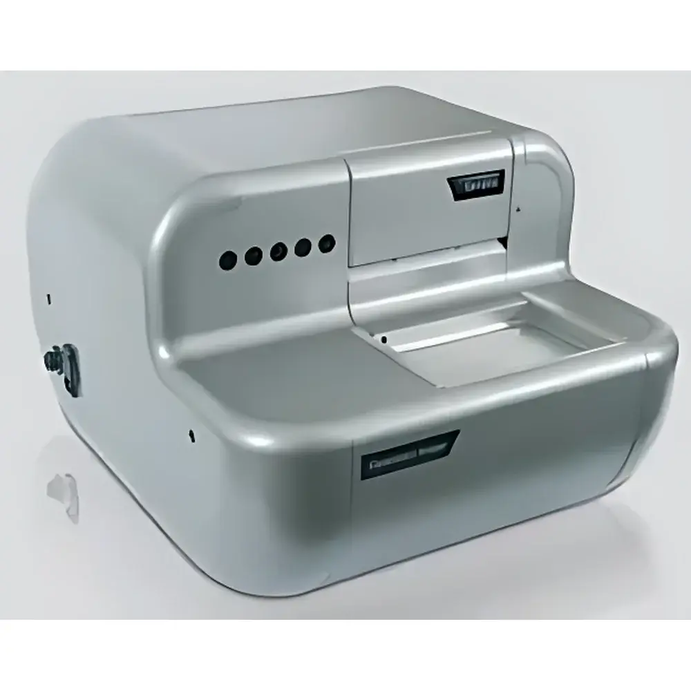
Molecular Devices CloneSelect Imager Cell Growth Analysis System
| Brand | Molecular Devices |
|---|---|
| Origin | United Kingdom |
| Manufacturer | Molecular Devices |
| Product Type | Imported Instrument |
| Model | CloneSelect Imager |
| Detection Principle | Label-Free Brightfield Imaging |
| Throughput | 96-Well Plate in 90 s |
| Application Focus | Monoclonal Verification, Clonogenic Assay, Cell Confluence & Proliferation Kinetics, Unlabeled Live-Cell Monitoring |
Overview
The Molecular Devices CloneSelect Imager Cell Growth Analysis System is a dedicated, label-free brightfield imaging platform engineered for objective, quantitative, and longitudinal monitoring of adherent and suspension cell cultures in standard microplates. Unlike fluorescence- or dye-based methods—which introduce cytotoxic stress, perturb physiological states, or require fixation—the CloneSelect Imager captures high-contrast, phase-enhanced brightfield images directly from live cells without exogenous labeling. Its core measurement principle relies on optical density contrast between cellular biomass and background substrate, enabling robust confluence estimation, single-cell-derived colony detection, and proliferation kinetics derivation across all wells simultaneously. Designed specifically for upstream bioprocess development and cell line engineering workflows, the system delivers time-resolved, plate-wide data essential for monoclonal verification, clonogenic assay quantification, culture condition optimization, and stability assessment—fully compatible with GLP-compliant documentation requirements.
Key Features
- Label-free, non-invasive brightfield imaging—eliminates phototoxicity, metabolic interference, and reagent costs associated with MTT, Calcein-AM, or Hoechst staining
- Full-plate acquisition in ≤90 seconds per 96-well plate, supporting multi-timepoint kinetic studies with minimal user intervention
- Automated confluence calculation per well using proprietary image segmentation algorithms trained on diverse morphologies (epithelial, fibroblastic, hybridoma, CHO, HEK293, iPSC-derived lines)
- “Loci of Growth” module for retrospective monoclonality verification: identifies wells containing a single origin of proliferation and reconstructs clonal lineage via temporal image stacks
- Integrated colony detection and sizing for semi-solid or soft-agar assays—quantifies colony count, area, circularity, and growth rate under compound treatment or media variation
- Native compatibility with robotic liquid handlers and incubator-integrated workflows via standard RS-232 and Ethernet interfaces
Sample Compatibility & Compliance
The CloneSelect Imager supports standard tissue-culture-treated 6-, 24-, 48-, and 96-well plates—including black-walled, clear-bottom, and ultra-low attachment formats—for both adherent monolayers and post-sedimentation suspension cultures (e.g., hybridomas, lymphocytes). It complies with ISO/IEC 17025 calibration traceability frameworks when operated within validated SOPs. Data integrity meets FDA 21 CFR Part 11 requirements through built-in audit trails, electronic signatures, and immutable storage of raw images, analysis parameters, and export logs. The system has been deployed in GMP-aligned cell line development suites at multiple global biopharmaceutical manufacturers and is routinely referenced in USP and ICH Q5D-compliant comparability protocols.
Software & Data Management
Acquisition and analysis are managed through CloneSelect Software v5.x—a Windows-based application featuring modular workflow templates (Monoclonality Check, Clonogenic Assay, Confluence Kinetics, Viability Screening). All images are stored in TIFF format with embedded EXIF metadata (timestamp, plate ID, exposure settings, focus position). Quantitative outputs—including confluence %, cell count estimates, colony metrics, and growth rate derivatives—are exportable to CSV, Excel, or LIMS-compatible XML. Batch processing supports plate-to-plate normalization, outlier rejection based on user-defined thresholds, and automated PDF report generation with embedded thumbnails, growth curves, and statistical summaries (mean ± SD, coefficient of variation). Audit trail records capture operator ID, timestamp, parameter edits, and result approvals—fully compliant with ALCOA+ data governance principles.
Applications
- Monoclonal Verification: Confirms clonal derivation by tracing proliferative origin across serial timepoints; satisfies regulatory expectations for clonality evidence in IND submissions
- Clonogenic Assays: Quantifies colony-forming efficiency (CFE) and drug-induced inhibition in oncology or toxicology screening, with dynamic area expansion metrics
- Culture Optimization: Accelerates serum-free or chemically defined media development by rapidly identifying conditions that maximize confluence rate and morphological uniformity
- Cell Line Stability Monitoring: Tracks long-term growth consistency post-thaw or after subcloning, detecting drift in doubling time or confluence saturation profiles
- ClonePix Integration: Validates and prioritizes clones isolated by ClonePix systems through objective growth trajectory comparison and expression correlation (when paired with secreted protein assays)
FAQ
Does the CloneSelect Imager require fluorescent dyes or cell labeling?
No—it uses label-free brightfield imaging optimized for native cellular contrast, eliminating cytotoxicity, assay variability, and regulatory concerns associated with exogenous labels.
Can it distinguish single cells from small clusters during early monoclonality assessment?
Yes—the “Loci of Growth” algorithm detects the spatial origin of proliferation and distinguishes true single-cell-derived colonies from aggregated artifacts when imaging begins within 24–48 hours post-seeding.
Is the system compatible with incubators or automated liquid handling platforms?
Yes—it supports integration via TTL triggers, RS-232 commands, and network-based API calls, and has been validated with major vendors including Hamilton STAR, Tecan Freedom EVO, and Binder C156 incubators.
How is data integrity ensured for regulatory submissions?
All image acquisitions, analysis parameters, and results are logged with timestamps, user IDs, and cryptographic hashes; full audit trails and electronic signature support align with 21 CFR Part 11 and Annex 11 requirements.
What plate formats and cell types have been empirically validated?
The system is validated for 6–96-well TC-treated plates and supports adherent lines (CHO-S, HEK293, A549), suspension-adapted lines (CHO-DG44, SP2/0), and primary cells (fibroblasts, MSCs) following standardized sedimentation protocols.
Related Products


