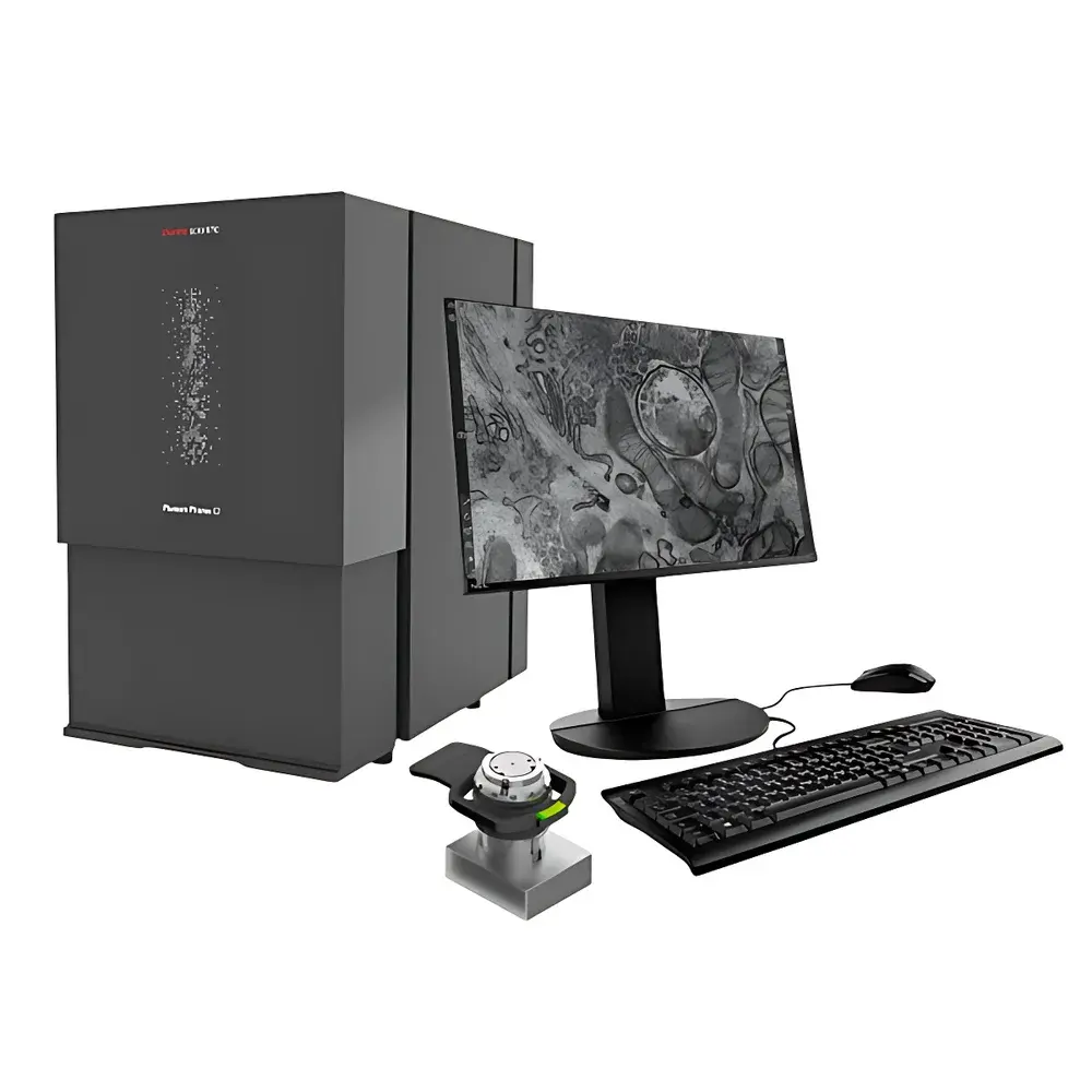
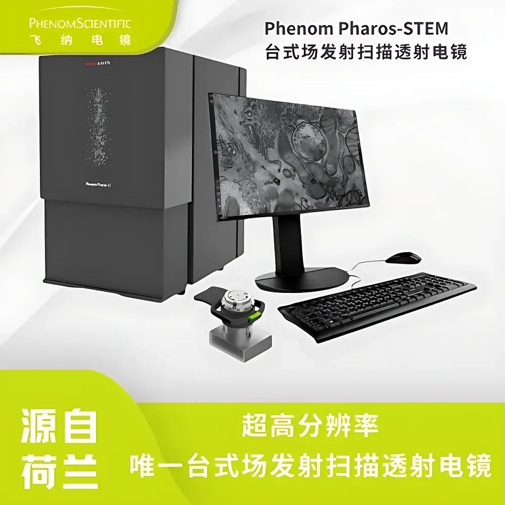
Phenom Pharos STEM Desktop Scanning Transmission Electron Microscope
| Brand | Phenom |
|---|---|
| Origin | Netherlands |
| Model | Pharos STEM |
| Accelerating Voltage | 20 kV |
| Resolution | ≤ 1 nm |
| Sample Holder | Standard 3 mm TEM grid clamp |
| Imaging Modes | Bright-Field (BF), Dark-Field (DF), High-Angle Annular Dark-Field (HAADF), User-Defined Detector Segmentation |
| Vacuum Levels | 0.1, 10, and 60 Pa |
| Imaging Time (Sample-to-Image) | < 40 s |
| Working Distance | Fixed for optimal STEM performance |
| Detector Configuration | 11-segment annular STEM detector with fully configurable quadrant/group selection |
Overview
The Phenom Pharos STEM is a purpose-engineered desktop scanning transmission electron microscope (STEM) designed for life science laboratories requiring high-resolution ultrastructural imaging without the infrastructure, expertise, or operational overhead of conventional high-voltage TEM systems. Unlike traditional transmission electron microscopes that rely on thermionic or lanthanum hexaboride (LaB₆) sources and complex column alignment, the Pharos STEM integrates a cold field-emission gun (FEG) operating at a fixed 20 kV accelerating voltage. This low-voltage configuration enables high-contrast imaging of beam-sensitive biological specimens—including unstained cryo-sections, exosomes, viral particles, and isolated macromolecular complexes—while minimizing radiation-induced damage and charging artifacts. The system operates in true scanning transmission mode: a focused electron probe scans across a thin specimen, and transmitted electrons are collected by an annular segmented detector array positioned below the sample. This architecture supports simultaneous acquisition of multiple contrast mechanisms—BF, DF, and HAADF—within a single scan, enabling quantitative structural correlation without mechanical reconfiguration.
Key Features
- Sub-nanometer resolution imaging: Achieves ≤ 1 nm spatial resolution under optimized conditions using a high-brightness cold FEG source, sufficient to resolve ribosomes, mitochondrial cristae, microtubule doublets, and synaptic vesicles in diagnostic pathology samples.
- 20 kV low-voltage STEM platform: Delivers superior mass-thickness contrast for organic materials while reducing ionization damage and minimizing specimen drift—critical for imaging hydrated or lightly fixed biological sections without heavy-metal staining.
- Integrated 11-segment annular detector: Enables real-time selection and combination of BF, DF, and HAADF signals; users may define custom detector groupings (e.g., inner/outer ring ratios, quadrant-specific integration) to emphasize specific scattering angles relevant to density gradients or crystallinity.
- TEM-grid compatible workflow: Accepts standard 3 mm diameter copper or nickel TEM grids via a precision-machined, kinematically aligned sample clamp—ensuring reproducible positioning and eliminating manual grid handling errors.
- Rapid vacuum cycle & one-click operation: Achieves operational vacuum (< 0.1 Pa) in under 15 seconds; full image acquisition—from grid loading to digital output—completes within 40 seconds, supporting high-throughput screening in clinical or teaching environments.
- Compact, vibration- and EMI-insensitive design: Fully self-shielded electron optics and passive damping enable stable operation on standard laboratory benches—even on upper floors—without dedicated foundations or Faraday cages.
Sample Compatibility & Compliance
The Pharos STEM is validated for use with ultrathin biological specimens prepared by conventional ultramicrotomy (50–100 nm sections), plunge-freezing followed by cryo-sectioning, and negative-stain or freeze-drying protocols. It accommodates standard 3 mm TEM grids (copper, nickel, gold, or silicon nitride membranes) without adapter modification. While not intended for routine high-resolution atomic-scale crystallography, its performance aligns with ASTM E1558 (Standard Guide for Transmission Electron Microscopy of Biological Specimens) and supports GLP-compliant documentation workflows when paired with Phenom’s audit-trail-enabled software. All imaging parameters—including kV, dwell time, pixel resolution, detector configuration, and vacuum log—are timestamped and exportable in vendor-neutral formats (TIFF, CSV) for regulatory submission.
Software & Data Management
Acquisition and analysis are managed through Phenom’s proprietary Phenom Desktop Software v5.x, which provides an intuitive, icon-driven interface compliant with ISO/IEC 17025 documentation requirements. The software includes built-in calibration routines traceable to NIST-certified grating standards, automatic contrast optimization algorithms, and batch-processing tools for multi-grid screening. Raw STEM data retains full detector segmentation metadata, enabling post-acquisition recombination of BF/DF/HAADF channels. Export options include TIFF (16-bit), PNG, and HDF5 for third-party analysis (e.g., ImageJ/Fiji, MATLAB, Python-based scikit-image pipelines). Audit logs record user ID, session start/end time, parameter changes, and image export events—fully satisfying FDA 21 CFR Part 11 requirements for electronic records when deployed with network authentication and role-based access control.
Applications
- Diagnostic pathology: Rapid screening of tissue ultrastructure at ×10,000–×50,000 magnification; visualization of endothelial fenestrations, podocyte foot processes, and myofibrillar organization across fields of view up to 200× larger than conventional TEM, significantly accelerating triage of renal, cardiac, or neuromuscular biopsies.
- Extracellular vesicle & viral characterization: High-contrast 3D-like topography of exosomes (30–150 nm), AAV capsids (~25 nm), tobacco mosaic virus rods (~18 nm diameter), and plasmid DNA condensates—without osmium tetroxide or uranyl acetate staining.
- Unstained biomolecular imaging: Direct observation of protein aggregates, amyloid fibrils, and lipid droplets in native-state preparations, avoiding dehydration-induced collapse and metal precipitate artifacts inherent to conventional TEM staining protocols.
- Teaching & core facility deployment: Designed for student-accessible operation with guided workflows, pre-set acquisition templates, and integrated safety interlocks—reducing training time and increasing instrument utilization in shared-resource environments.
FAQ
What sample thickness is optimal for Pharos STEM imaging?
For reliable BF/HAADF contrast at 20 kV, biological sections should be 50–80 nm thick—compatible with standard diamond-knife ultramicrotomy. Thicker sections (>120 nm) may yield excessive multiple scattering, reducing HAADF signal fidelity.
Can the Pharos STEM perform energy-dispersive X-ray spectroscopy (EDS)?
No. The Pharos STEM is optimized exclusively for STEM imaging; it does not integrate an EDS detector or support X-ray microanalysis. Elemental mapping requires coupling with a separate SEM-EDS or TEM-EDS platform.
Is cryo-transfer supported?
The system does not include a cryo-stage or transfer mechanism. Ambient-temperature imaging of air-dried, critical-point-dried, or room-temperature cryo-sections is supported; full cryo-STEM functionality requires a dedicated high-end TEM.
How is resolution verified and maintained?
Resolution validation uses certified gold-on-carbon resolution test gratings (NIST SRM 2051); daily quick-checks employ polycrystalline aluminum oxide diffraction patterns. No user-serviceable optical alignment is required—the column is factory-aligned and sealed.
What maintenance is required?
Annual filament replacement and vacuum pump oil change are the only scheduled interventions. The FEG source lifetime exceeds 1,500 hours; no column cleaning, aperture replacement, or lens calibration is needed during normal operation.


