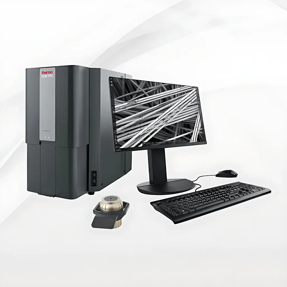
Phenom Pro_2 Desktop Scanning Electron Microscope for Life Sciences
| Brand | Phenom |
|---|---|
| Origin | Netherlands |
| Manufacturer Type | Original Equipment Manufacturer (OEM) |
| Origin Category | Imported |
| Model | Phenom Pro_2 |
| Instrument Type | Benchtop SEM |
| Electron Source | Cerium Hexaboride (CeB₆) |
| Secondary Electron Resolution | 6 nm |
| Maximum Magnification | 350,000× |
| Accelerating Voltage Range | 4.8–20.5 kV |
| Backscattered Electron Resolution | 8 nm |
Overview
The Phenom Pro_2 is a high-performance benchtop scanning electron microscope (SEM) engineered specifically for life science laboratories requiring rapid, reliable, and high-fidelity ultrastructural imaging without the operational complexity or infrastructure demands of floor-standing systems. Utilizing a thermionic cerium hexaboride (CeB₆) electron source, the Phenom Pro_2 delivers stable beam current and superior signal-to-noise ratio—critical for imaging delicate biological specimens such as fixed tissues, cryo-prepared cells, biofilms, coral microbial surfaces, and other beam-sensitive samples. Its compact design integrates vacuum generation, electron optics, and real-time imaging into a single footprint (<0.5 m²), enabling deployment in standard biosafety cabinets, core imaging facilities, or teaching labs. Unlike conventional SEMs requiring liquid nitrogen cooling or high-vacuum pumping stages, the Phenom Pro_2 employs a fully integrated turbomolecular pump system with automated pressure management, ensuring consistent chamber conditions across variable sample types and preparation protocols.
Key Features
- Real-time hybrid imaging mode: Simultaneous acquisition and on-the-fly blending of secondary electron (SE) and backscattered electron (BSE) signals—enabling topographic contrast (SE) and atomic number contrast (BSE) in a single view.
- Enhanced resolution architecture: Sixth-generation optical alignment and detector optimization deliver 20% improved SE resolution (6 nm at 10 kV) and 8 nm BSE resolution—validated per ISO 16700:2016 guidelines for SEM resolution measurement.
- Intuitive workflow interface: Touchscreen-driven acquisition software with one-click auto-focus, auto-stigmation, and stage navigation—requiring no prior SEM training for routine operation.
- Bio-optimized beam control: Adjustable accelerating voltage (4.8–20.5 kV) with low-kV capability for charge mitigation on non-conductive biological samples; optional low-vacuum mode supports hydrated or partially coated specimens.
- Modular expandability: Native compatibility with EDS (energy-dispersive X-ray spectroscopy) detectors for elemental mapping—enabling seamless upgrade to Phenom ProX configuration without hardware replacement.
Sample Compatibility & Compliance
The Phenom Pro_2 accommodates a broad range of life science specimen formats—including resin-embedded tissue sections, sputter-coated cell monolayers, freeze-dried microbial colonies, and uncoated botanical or marine biostructures (e.g., coral endolithic bacterial surfaces). Sample holders accept standard 12 mm pin stubs or proprietary multi-well specimen cups, supporting parallel analysis of up to 16 samples per session. All vacuum and imaging parameters comply with IEC 61000-6-3 (EMC emissions) and IEC 61000-6-2 (immunity), and the system’s firmware architecture supports audit trails and user-access logging required under GLP and ISO/IEC 17025-accredited laboratory environments. While not FDA-cleared as a diagnostic device, its imaging output meets ASTM E1558-21 standards for SEM-based morphological characterization in preclinical research.
Software & Data Management
Acquisition and analysis are managed via Phenom Desktop Software v5.x—a Windows-based platform supporting DICOM-compliant image export, TIFF/PNG batch saving with embedded metadata (kV, WD, dwell time, detector gain), and histogram-based contrast optimization. The software includes built-in measurement tools (line profiles, particle counting, area fraction analysis) compliant with ISO 13322-2 for particle size distribution reporting. For regulated environments, optional 21 CFR Part 11 compliance packages provide electronic signature validation, role-based permissions, and immutable audit logs—fully traceable to individual user sessions and instrument parameter changes.
Applications
- Ultrastructural analysis of mammalian and plant cell organelles, extracellular matrix architecture, and pathogen-host interface morphology.
- Characterization of biofilm topography, microbial colony morphology, and calcified biomineral structures (e.g., coral skeletal porosity and endolithic bacterial colonization patterns).
- Quality control of biomaterial scaffolds, electrospun nanofibers, and lyophilized pharmaceutical formulations.
- Teaching and demonstration of electron microscopy principles in undergraduate and graduate life science curricula.
- Correlative workflows: Integration with light microscopy (LM) and fluorescence data via coordinate-mapped stage navigation for multimodal structural-functional correlation.
FAQ
Is the Phenom Pro_2 suitable for imaging unstained, non-conductive biological samples?
Yes—its low-kV imaging capability (down to 4.8 kV), combined with optional low-vacuum mode and charge compensation algorithms, enables high-contrast imaging of lightly fixed or critical-point-dried specimens without metal coating.
Can the system be validated for use in ISO 17025-accredited testing laboratories?
Yes—the instrument supports full calibration documentation, traceable to NIST-traceable reference standards, and its software audit trail functionality satisfies clause 7.7 of ISO/IEC 17025:2017 for result integrity verification.
What is the typical turnaround time from installation to first image?
Under standard lab conditions (23°C ±2°C, <60% RH, vibration-isolated bench), users achieve operational readiness—including vacuum stabilization and system calibration—in under 15 minutes post-power-on.
Does the Phenom Pro_2 support automated particle analysis for microbiology applications?
Yes—integrated particle analysis modules enable threshold-based segmentation, size distribution histograms, and shape factor quantification (circularity, aspect ratio), all exportable in CSV format for statistical validation per ISO 9276-2.