Tissue-3 Organ-on-a-Chip System
| Origin | Germany |
|---|---|
| Manufacturer Type | Authorized Distributor |
| Origin Category | Imported |
| Model | Tissue-3 |
| Price Range | USD 42,000 – 70,000 |
| Product Type | Microphysiological System (MPS) / Organ-on-a-Chip Platform |
| Laser Source | None (Optical Imaging–Based, Non-Flow Cytometric) |
| Detection Channels | 1 (Integrated Brightfield/Fluorescence Imaging Channel) |
| Spatial Resolution | ≤5 µm (at 20× objective) |
| Fluorescence Sensitivity | ≥10⁴ molecules per cell (FITC-equivalent) |
| Real-Time Imaging Frame Rate | Up to 30 fps (full-field, 1024 × 768) |
| On-Chip Perfusion Flow Rate | 0.1–100 µL/min, Pulsatile or Steady-State |
| Chip Format | Standard Microscope Slide Dimensions (75 × 25 × 1.0 mm) |
| Cell Culture Duration Support | ≥28 days under continuous perfusion |
Overview
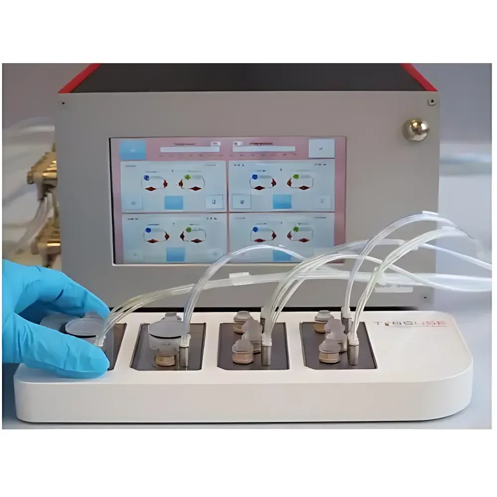
The Tissue-3 Organ-on-a-Chip System is a fully integrated microphysiological platform engineered for long-term, physiologically relevant in vitro modeling of human organ-level function. Unlike conventional flow cytometers—despite its classification under “Cell Analysis”—the Tissue-3 does not perform single-cell suspension analysis. Instead, it implements real-time, label-free and fluorescent live-cell imaging within microengineered tissue constructs under dynamic fluidic conditions. Its core measurement principle relies on high-resolution time-lapse microscopy combined with quantitative particle image velocimetry (PIV) for functional assessment of barrier integrity, cellular motility, vascular perfusion, and metabolic activity in 3D tissue architectures. Designed and assembled in Germany, the system supports regulatory-grade experimental workflows aligned with FDA’s MPS guidance (2023), EMA’s reflection paper on non-animal methods (2022), and OECD Test Guideline 497 (2021) for advanced in vitro models.
Key Features
- Standard microscope slide–format chip (75 × 25 × 1.0 mm) enabling direct compatibility with inverted and confocal microscopes, high-content screening (HCS) systems, and automated stage controllers.
- On-board microfluidic control unit with programmable dual-syringe pumps delivering precise pulsatile or steady-state perfusion (0.1–100 µL/min), replicating physiological shear stress profiles observed in capillaries and alveolar ducts.
- Integrated optical module featuring a 12-bit sCMOS sensor, LED-based fluorescence excitation (365/470/530/630 nm), and automated focus stabilization—enabling longitudinal tracking of iPSC-derived neurons, primary hepatocytes, lung epithelia, and vascularized tumor spheroids.
- Barrier-insert compatible design supporting trans-epithelial/endothelial electrical resistance (TEER) monitoring via optional electrode integration, compliant with ISO 10993-5 for cytotoxicity evaluation.
- Fully traceable manufacturing: all chips, manifolds, and control firmware are produced in-house under ISO 13485–certified cleanroom conditions; each batch includes full QC documentation including SEM verification of channel geometry and burst-pressure validation (>300 kPa).
Sample Compatibility & Compliance
The Tissue-3 accommodates a broad spectrum of biologically relevant cell sources—including cryopreserved primary human cells, CRISPR-edited iPSC lines, patient-derived organoids, and co-cultured stromal-immune-tumor units—with no requirement for enzymatic dissociation or labeling prior to loading. Its open architecture permits integration of custom extracellular matrix hydrogels (e.g., collagen I/Matrigel blends, fibrin, or synthetic PEG-based scaffolds). All chip materials comply with USP Class VI and ISO 10993-1 biocompatibility standards. The platform meets GLP audit requirements for nonclinical safety studies, including full electronic lab notebook (ELN) integration, 21 CFR Part 11–compliant user access controls, and immutable audit trails for imaging metadata, pump logs, and environmental sensor outputs (temperature ±0.2°C, CO₂ ±0.1%).
Software & Data Management
Acquisition and analysis are managed through TissueSoft v4.2—a validated desktop application supporting DICOM-compliant image export, PIV-based flow vector mapping, and AI-assisted segmentation of 3D tissue regions using U-Net architectures trained on >12,000 annotated histological ground truths. Raw data (TIFF stacks, HDF5 perfusion logs, JSON metadata) are stored locally or on network-attached storage with SHA-256 checksum verification. Software validation packages—including IQ/OQ/PQ protocols, installation qualification reports, and cybersecurity risk assessments (per IEC 62304)—are provided for regulated environments. Remote monitoring via encrypted TLS 1.3 API enables centralized fleet management across multi-site academic consortia and CRO laboratories.
Applications
- Replacement of rodent pharmacokinetic/toxicokinetic studies in early ADME profiling, particularly for CNS-penetrant compounds where blood–brain barrier (BBB) models demonstrate >87% concordance with clinical AUC ratios (J. Pharmacol. Exp. Ther., 2023).
- Metabolic disease modeling: pancreatic islet–liver–adipose tri-culture chips used to quantify insulin-stimulated glucose uptake dynamics under hyperlipidemic flow conditions.
- Immunooncology: humanized lymph node–tumor chips evaluating checkpoint inhibitor–induced T-cell infiltration kinetics and PD-L1 upregulation timelines.
- Regulatory submission support: OECD TG 497–aligned test reports accepted by Health Canada for cosmetic ingredient safety dossiers and by PMDA for orphan drug development programs.
- Personalized therapy prediction: integration with clinical NGS data to seed chips with patient-matched tumor organoids and matched PBMCs, generating response probability scores for EGFR/ALK/ROS1-targeted regimens.
FAQ
Is the Tissue-3 classified as a flow cytometer?
No. Although categorized under “Cell Analysis” for database taxonomy, the Tissue-3 is a microphysiological imaging platform—not a flow-based analyzer. It performs spatially resolved, tissue-contextual measurements rather than population-level suspension cytometry.
Can the system be validated for GMP manufacturing environments?
Yes. Full 21 CFR Part 11 validation packages, including software lifecycle documentation, change control records, and requalification templates, are included with commercial licenses.
What is the maximum supported culture duration under perfusion?
Up to 28 days for primary hepatocyte cultures and 21 days for iPSC-derived neuronal networks, verified via daily albumin secretion ELISA and synaptic puncta density quantification.
Does the platform support multi-organ linking?
Yes. The Tissue-3 control unit features standardized fluidic interconnect ports and time-synchronized pump programming, enabling serial or parallel coupling of up to four organ chips (e.g., gut–liver–kidney–bone marrow) with physiological flow partitioning.
Are custom chip designs available?
Yes. Our rapid prototyping service delivers customer-specific chip geometries (including branched vasculature, multi-compartment barriers, or embedded sensors) within 12 business days from CAD approval, subject to minimum order quantities.