MMI Cellcut Plus Live-Cell Laser Microdissection System
| Origin | Germany |
|---|---|
| Manufacturer Type | Authorized Distributor |
| Origin Category | Imported |
| Model | Cellcut Plus |
| Price | Upon Request |
Overview
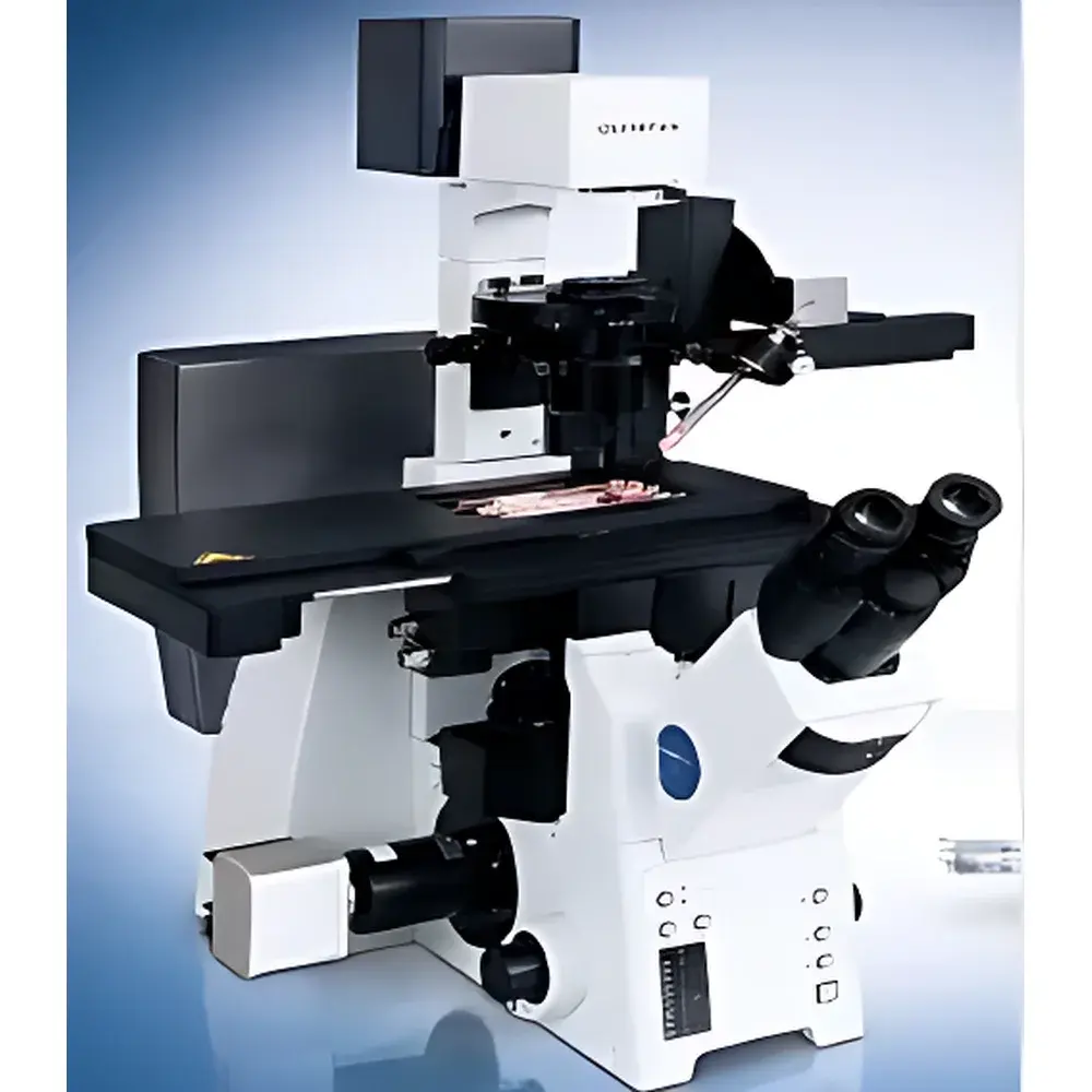
The MMI Cellcut Plus is a high-precision, live-cell compatible laser microdissection (LMD) system engineered for spatially resolved isolation of intact, viable cells and subcellular structures under optical microscopy guidance. Unlike conventional thermal LMD platforms that rely on infrared or UV lasers inducing localized ablation or membrane perforation, the Cellcut Plus employs a patented *cold ultraviolet (UV) solid-state laser* operating at 355 nm—delivering sub-micron beam diameter (<1 µm) with negligible photothermal damage. This enables non-destructive, contact-free cutting of delicate biological specimens—including adherent live cells in culture, frozen or FFPE tissue sections, and even plant tissues—while preserving RNA integrity, protein conformation, and cellular viability for downstream functional assays such as clonal expansion, single-cell RNA sequencing, and live imaging. Its core architecture integrates an inverted research-grade microscope (Olympus IX71 or Nikon Ti-S), motorized high-resolution XY stage, and real-time 3-chip CCD imaging for true-color visualization—ensuring morphological fidelity during selection and retrieval.
Key Features
- Patented cold UV laser (355 nm, solid-state) with <1 µm spot size—enabling chromosome-level dissection without thermal artifact or nucleic acid degradation.
- Membrane-based capture technology: Samples remain sandwiched between glass slide and thermoplastic film, eliminating direct mechanical contact, cross-contamination, or shear-induced stress during collection.
- Eppendorf tube cap interfaces exclusively with the film—not the specimen—ensuring sterility and preventing non-specific adhesion or carryover.
- Gentle, non-ablative separation: No laser irradiation required for transfer; captured regions are lifted intact via controlled film retraction, allowing real-time visual verification of cell morphology pre- and post-isolation.
- Optimized for variable hydration states: Compatible with air-dried, ethanol-fixed, cryosections, paraffin-embedded, and live-cell monolayers—no stringent dehydration protocols needed.
- True-color 3-chip CCD camera (RGB-separated sensors) delivers accurate spectral reproduction—critical for distinguishing subtle cytoplasmic/nuclear features, fluorescent markers, or histochemical stains in heterogeneous samples.
- Modular design supports future upgrade to suspension cell harvesting (e.g., flow-sorted or spheroid-derived cells) via optional fluidic integration.
Sample Compatibility & Compliance
The Cellcut Plus accommodates diverse biospecimen formats across translational and basic research workflows: formalin-fixed paraffin-embedded (FFPE) tissue sections (4–20 µm), OCT-embedded cryosections, fresh-frozen sections, live adherent cultures (e.g., primary neurons, epithelial monolayers, iPSC-derived organoids), and plant tissue sections. It complies with ISO 13485:2016 for medical device quality management systems and supports GLP/GMP-aligned documentation through audit-trail-enabled software logging. All hardware components—including laser source, stage controllers, and imaging modules—meet CE marking requirements for in vitro diagnostic (IVD) instrumentation under Directive 98/79/EC. The system is validated for compatibility with downstream molecular applications compliant with ISO/IEC 17025 and ASTM E2554 standards for nucleic acid integrity assessment.
Software & Data Management
The MMI CellCut Software Suite provides intuitive touchscreen-driven operation with integrated image annotation, region-of-interest (ROI) mapping, and automated coordinate registration between microscope field-of-view and stage position. Each dissection event is timestamped and logged with metadata including laser parameters (pulse energy, repetition rate), ROI coordinates, user ID, and acquisition settings—supporting full traceability per FDA 21 CFR Part 11 requirements when configured with electronic signature and audit trail modules. Export formats include TIFF, PNG, and OME-TIFF for interoperability with ImageJ/Fiji, QuPath, and commercial pathology platforms. Raw image data and log files are stored in structured directory hierarchies compliant with FAIR (Findable, Accessible, Interoperable, Reusable) principles, facilitating integration into institutional LIMS or ELN environments.
Applications
- Single-cell isolation for scRNA-seq, scATAC-seq, and multi-omics profiling from heterogeneous tumor microenvironments.
- Viable cell recovery from primary cultures for clonal expansion, functional assays, or transplantation studies.
- Region-specific harvesting of defined anatomical structures (e.g., hippocampal subfields, cortical layers, glomeruli) from brain or kidney sections.
- Chromosome microdissection for cytogenetic analysis, FISH probe generation, or CRISPR target validation.
- Plant cell-type-specific sampling (e.g., guard cells, trichomes, vascular bundles) without enzymatic digestion artifacts.
- Quality-controlled isolation of rare circulating tumor cells (CTCs) or disseminated tumor cells (DTCs) from spiked blood smears.
FAQ
Is the Cellcut Plus suitable for RNA-seq-ready sample preparation?
Yes—the cold UV laser minimizes RNA fragmentation, and the membrane-capture workflow avoids RNase exposure; users routinely achieve RIN >8.5 from FFPE and frozen sections.
Can I integrate fluorescence imaging for marker-guided dissection?
Yes—both Olympus IX71 and Nikon Ti-S configurations support modular fluorescence modules (DAPI/FITC/TRITC/Cy5), enabling immunofluorescence- or GFP-guided selection.
What maintenance does the UV laser require?
None—the diode-pumped solid-state laser has a rated lifetime exceeding 10,000 hours and no consumables or alignment procedures.
Does the system support batch processing of multiple slides?
Yes—up to three standard microscope slides can be loaded simultaneously and processed sequentially with automatic focus and ROI registration.
How is sterility maintained during live-cell harvesting?
Sterility is ensured by laminar-flow-compatible enclosure options, UV sterilization cycles, and the film-mediated transfer mechanism that prevents direct contact between tubes and biological material.
Related Products