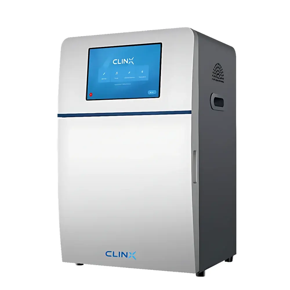
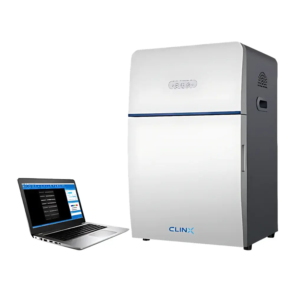
ChemiScope 6200 Multifunctional Chemiluminescence Imaging System
| Brand | CLINX |
|---|---|
| Origin | Shanghai, China |
| Model | ChemiScope 6200 |
| Instrument Type | Chemiluminescence Gel Imaging System |
| CCD Resolution | 6 MP |
| Sensor Cooling | −25 °C |
| Bit Depth | 16-bit (65,536 gray levels) |
| Pixel Binning Options | 1×1, 2×2, 3×3, 4×4 |
| Lens | F/0.95 motorized auto-focus lens |
| UV Transilluminator (optional) | 365 nm or 380 nm LED, 21 cm × 26 cm area |
| Blue Light Transilluminator (optional) | 470 nm LED, 21 cm × 26 cm |
| White Light Transilluminator (optional) | LED, 21 cm × 26 cm |
| Reflective Fluorescence Excitation (optional) | 365/470/530/630/660/730 nm LEDs |
| Emission Filters (optional) | 460/525/585/690/715/810 nm |
| Sample Stage | Dual-layer chemiluminescence stage with white LED reflectance illumination |
| Software | ChemiScope Image Acquisition & Analysis Suite |
| Data Interface | Dual USB 3.0 ports |
| Touchscreen | Optional 10.1″ industrial embedded panel PC |
Overview
The CLINX ChemiScope 6200 is a high-performance, multifunctional chemiluminescence and fluorescence imaging system engineered for quantitative molecular detection across Western blotting, nucleic acid electrophoresis, protein gel analysis, and bioluminescent reporter assays. It employs a thermoelectrically cooled 6-megapixel CCD sensor (−25 °C operating temperature) with true 16-bit dynamic range (65,536 gray levels), enabling low-light signal capture with high reproducibility and minimal thermal noise. The system operates on the principle of photon detection via intensified charge-coupled device imaging—optimized for both enzymatic chemiluminescence (e.g., horseradish peroxidase–luminol or alkaline phosphatase–CSPD systems) and direct fluorescence excitation/emission across UV–visible–NIR spectral bands. Its modular optical architecture supports interchangeable transillumination and reflective excitation sources, allowing seamless transition between ECL membrane imaging, ethidium bromide–stained DNA gels, SYBR Safe–labeled RNA, Cy5-labeled antibodies, and luciferase-based in vivo bioluminescence samples—all within a single platform compliant with GLP documentation requirements.
Key Features
- Thermoelectrically cooled 6 MP CCD camera with −25 °C stabilization, delivering low dark current and high quantum efficiency for sub-femtogram protein detection.
- F/0.95 motorized auto-focus lens with variable pixel binning (1×1 to 4×4) to balance resolution, sensitivity, and acquisition speed per experimental requirement.
- Dual-layer sample stage with integrated white LED reflectance illumination for uniform chemiluminescence signal capture from blot membranes without external darkroom dependency.
- Intelligent light-source recognition: automatically detects installed transilluminator or excitation module (UV/Blue/White/fluorescence) and configures optimal exposure parameters and filter wheel position.
- Long-life LED light sources (>30,000 h lifetime), with >90% irradiance uniformity across 21 cm × 26 cm active area—eliminating hotspots and improving quantitation linearity.
- Optional 10.1″ embedded industrial touchscreen interface with multi-touch support; also compatible with external Windows-based workstations via USB 3.0.
Sample Compatibility & Compliance
The ChemiScope 6200 accommodates standard and mini-format polyacrylamide and agarose gels, nitrocellulose/PVDF membranes, microplates (up to 96-well), and tissue sections. It supports all major nucleic acid stains (Ethidium Bromide, SYBR Gold/Green/Safe, GelStar, GelSignal Red/Green) and protein visualization methods (Coomassie Brilliant Blue, silver stain, fluorescent dyes, and label-free total protein staining). For regulatory environments, the system’s software enforces audit-trail logging per FDA 21 CFR Part 11 requirements—including user authentication, timestamped image metadata, parameter change history, and electronic signature-capable PDF report generation. All optical components comply with IEC 61000-4 electromagnetic compatibility standards and are housed in an interlocked UV-shielded enclosure meeting EN 60825-1 laser safety classification guidelines where applicable.
Software & Data Management
The ChemiScope Image Acquisition & Analysis Suite provides a validated, role-based workflow environment. Core modules include: (1) Acquisition—featuring preconfigured templates for WB, DNA gel, fluorescence, and bioluminescence; manual/auto exposure control; real-time histogram preview; and lossless TIFF export; (2) Quantitative Analysis—automated lane and band detection with adjustable sensitivity thresholds, background subtraction using rolling-ball algorithm, molecular weight calibration via ladder fitting (linear/logarithmic interpolation), and integrated densitometry with % volume and integrated intensity metrics; (3) Account Management—multi-tier permission structure (Admin, Supervisor, User), full action logging, and encrypted local database storage. Export formats include Excel (.xlsx) for raw data, annotated TIFF/PNG for publication, and PDF reports embedding metadata, operator ID, acquisition parameters, and analysis settings—fully traceable for ISO/IEC 17025 or CAP-accredited labs.
Applications
- Western Blot chemiluminescence imaging using ECL, ECL Plus, or ChemiSignal® Plus reagents—capable of detecting <10 fmol target protein per band under optimized conditions.
- Northern and Southern blot analysis with DIG- or radioisotope-free detection strategies.
- SDS-PAGE and native PAGE gel documentation for Coomassie- and silver-stained proteins, including post-electrophoretic fluorescence labeling.
- Agarose and polyacrylamide nucleic acid gel imaging across UV-excitable (EtBr), blue-light–compatible (GelSignal Green), and visible-light–safe (SYBR Safe) dyes.
- Fluorescence multiplexing using Cy2/Cy3/Cy5, Alexa Fluor, GFP/RFP fusion proteins, and near-infrared probes (e.g., IRDye 800CW) with spectral unmixing support via sequential filter acquisition.
- Bioluminescent reporter assays in cell lysates or live animal imaging (ex vivo tissue slices), leveraging low-background detection and extended integration times up to 60 minutes.
FAQ
What is the maximum exposure time supported for chemiluminescent imaging?
The system supports exposure durations from 10 ms to 60 minutes, adjustable in 1-ms increments for precise signal optimization.
Does the software support batch processing of multiple images with identical analysis parameters?
Yes—batch mode enables automated lane detection, background correction, and densitometric quantification across folders of TIFF or PNG files using saved analysis profiles.
Can the ChemiScope 6200 be validated for use in GMP-regulated environments?
The system supports IQ/OQ documentation packages, 21 CFR Part 11-compliant audit trails, electronic signatures, and configurable user roles—enabling qualification per ASTM E2500 and EU Annex 11 guidelines.
Is third-party filter or lens compatibility supported?
Only CLINX-certified optical components are guaranteed for performance and safety compliance; non-OEM filters may compromise spectral fidelity or violate UV shielding certifications.
How is calibration maintained across long-term usage?
The system includes built-in flat-field correction routines and optional NIST-traceable reference standards for daily intensity verification and grayscale linearity validation.