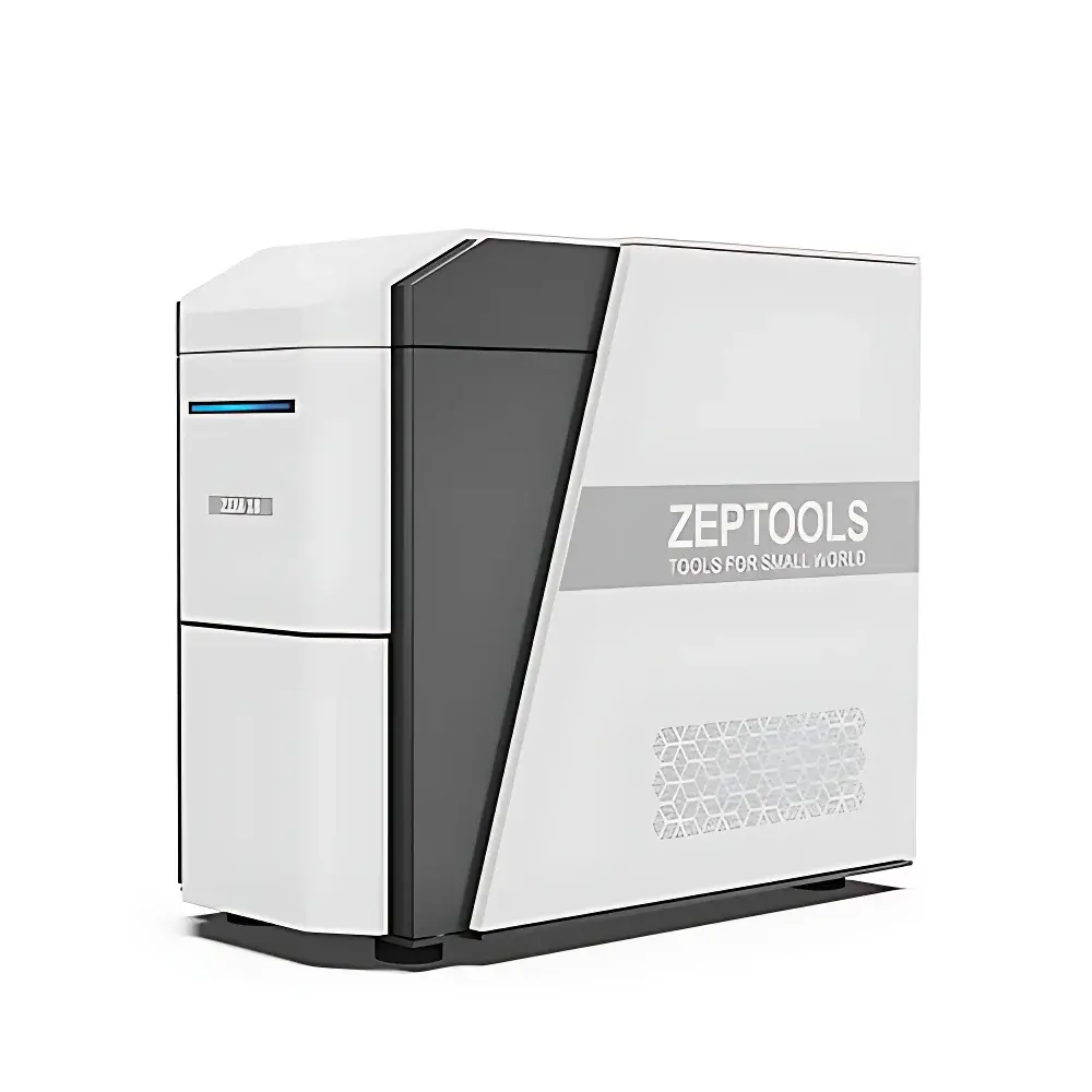
ZEPTOOLS ZEM18 Desktop Scanning Electron Microscope
| Brand | ZEPTOOLS |
|---|---|
| Origin | Anhui, China |
| Model | ZEM18 |
| Instrument Type | Desktop SEM |
| Electron Source | Pre-aligned Tungsten Filament |
| Acceleration Voltage | 3–18 kV (1 kV step) |
| Max. Magnification | 200,000× |
| Resolution | <6 nm @ 15 kV, high vacuum mode |
| Standard Detectors | Secondary Electron (SE), Backscattered Electron (BSE) |
| Vacuum System | High vacuum (≤5×10⁻³ Pa) |
| Sample Exchange Time | ≤90 s |
| Stage Options | 2-axis mechanical stage (standard) |
| Navigation | Integrated optical navigation camera |
| Optional Add-ons | EDS detector, in-situ heating/cooling stages (TEC), deceleration stage |
Overview
The ZEPTOOLS ZEM18 is a compact, high-performance desktop scanning electron microscope engineered for routine microstructural characterization in academic laboratories, quality control environments, and industrial R&D settings. Utilizing thermionic emission from a pre-aligned tungsten filament, the ZEM18 delivers stable electron beam generation with minimal maintenance overhead. Its optimized column design and integrated electromagnetic lens system enable sub-6 nm resolution imaging at 15 kV under high vacuum conditions—meeting ASTM E1558 and ISO 16700 requirements for SEM performance verification. Unlike conventional floor-standing systems, the ZEM18 integrates vacuum pumping, beam control, signal detection, and image acquisition into a single benchtop enclosure, eliminating the need for dedicated instrument rooms or external vibration isolation. The system operates within a controlled high-vacuum environment (base pressure ≤5×10⁻³ Pa), with an optional low-vacuum mode (1–60 Pa) supporting semi-conductive or mildly outgassing samples without metal coating.
Key Features
- Rapid vacuum cycling: Achieves operational vacuum in ≤90 seconds via a dual-stage turbomolecular pump system, significantly reducing turnaround time between samples.
- Flexible acceleration voltage control: Continuously adjustable from 3 kV to 18 kV in 1 kV increments—enabling optimization of surface sensitivity (low-kV imaging) and penetration depth (high-kV analysis) across diverse material classes.
- Dual-detector architecture: Standard integration of both secondary electron (SE) and backscattered electron (BSE) detectors provides simultaneous topographic and atomic number contrast imaging, essential for phase identification and grain boundary analysis.
- Optical navigation system: Built-in coaxial optical camera enables real-time macro-to-micro sample positioning with sub-millimeter accuracy, streamlining region-of-interest selection prior to SEM imaging.
- Modular stage configuration: Base model includes a robust 2-axis mechanical stage; upgradeable to a 3-axis motorized stage with ±25 mm X/Y travel and 10 mm Z lift for extended mapping and tilt-series acquisition.
- In-situ compatibility: Designed with standardized flange interfaces (CF35/CF40) to support third-party and ZEPTOOLS-developed in-situ accessories—including resistive heating stages (up to 1000 °C), thermoelectric cooling stages (–180 °C to +150 °C), and deceleration-based low-energy surface analysis modules.
Sample Compatibility & Compliance
The ZEM18 accommodates standard 32 mm diameter stub-mounted specimens up to 40 mm in height. Its high-vacuum operation requires conductive coating (e.g., Au/Pd sputtering) for non-conductive samples; however, the optional low-vacuum mode permits direct imaging of hydrated polymers, biological tissues, and uncoated ceramics at pressures up to 60 Pa. All hardware and software components comply with IEC 61000-6-3 (EMC emissions) and IEC 61010-1 (safety requirements for electrical equipment). Data integrity protocols align with GLP and GMP documentation standards, including timestamped parameter logging, user-access-controlled method files, and audit-trail-enabled session records—facilitating regulatory submissions under FDA 21 CFR Part 11 where electronic signatures are required.
Software & Data Management
Acquisition and analysis are managed through ZEPTOOLS’ proprietary SEM Suite v4.x—a Windows-based platform supporting real-time image preview, multi-frame averaging, drift correction, and automated focus/stigmation routines. The software implements lossless TIFF export with embedded metadata (kV, WD, dwell time, detector gain), compatible with open-format analysis tools such as ImageJ/Fiji, DigitalMicrograph, and MATLAB. Batch acquisition workflows support grid-based automated imaging, particle size distribution analysis (PSD), and EDS spectral library matching (when EDS module is installed). All session data—including raw images, parameter sets, and annotation layers—are stored in a relational SQLite database with configurable backup intervals and network share synchronization.
Applications
The ZEM18 serves as a primary characterization tool in materials science labs evaluating battery electrode morphology, catalyst nanoparticle dispersion, and fracture surface topography. In semiconductor process development, it supports failure analysis of lithographic patterns, interconnect void detection, and cross-sectional metrology of thin-film stacks. Life science users apply it for imaging freeze-fractured membranes, mineralized tissue sections, and microporous drug delivery carriers. Its rapid workflow and intuitive interface also make it suitable for undergraduate teaching laboratories, where students perform hands-on training in vacuum fundamentals, electron-sample interactions, and digital image interpretation—without requiring dedicated facility infrastructure.
FAQ
What is the typical resolution specification, and under what conditions is it measured?
Resolution is specified as <6 nm at 15 kV, measured on a clean gold-on-carbon reference standard under high-vacuum conditions using the SE detector and optimal working distance (WD = 10 mm).
Is EDS elemental analysis supported natively?
EDS functionality is available as a factory-installed option with full software integration, including spectrum acquisition, qualitative peak identification (based on NIST Standard Reference Database), and semi-quantitative ZAF matrix corrections.
Can the ZEM18 accommodate large or irregularly shaped samples?
Maximum sample height is 40 mm; lateral dimensions are constrained by the chamber’s internal diameter (120 mm). Custom stub adapters and removable pole-piece inserts allow limited accommodation of oversized geometries, subject to clearance verification during installation.
Does the system support automated stage mapping and stitching?
Yes—SEM Suite v4.x includes mosaic acquisition with auto-overlap detection, distortion correction, and seamless image stitching for areas exceeding single-field-of-view dimensions.
What vacuum pump maintenance schedule is recommended?
The turbomolecular pump requires annual bearing inspection and oil change in the backing pump; full service intervals are documented in the Maintenance Manual and tracked via built-in pump runtime counters.


