Auniontech DOIT Fourier Light-Field Microscopy Camera Module for 3D Real-Time Digital Imaging
| Brand | Auniontech |
|---|---|
| Origin | Shanghai, China |
| Manufacturer Type | Authorized Distributor |
| Product Category | Domestic |
| Model | DOIT Light-Field Microscopy Imaging Module |
| Price | Upon Request |
| Depth of Field | 160 µm |
Overview
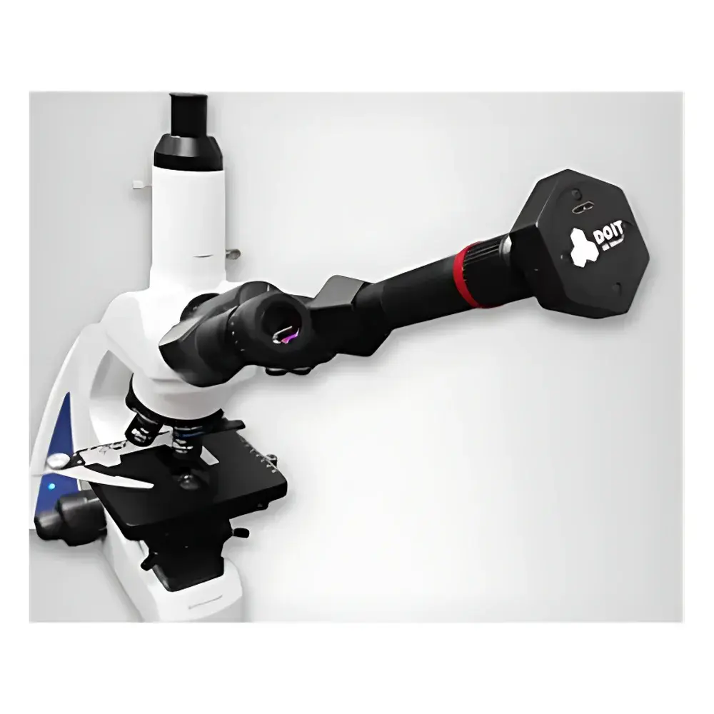
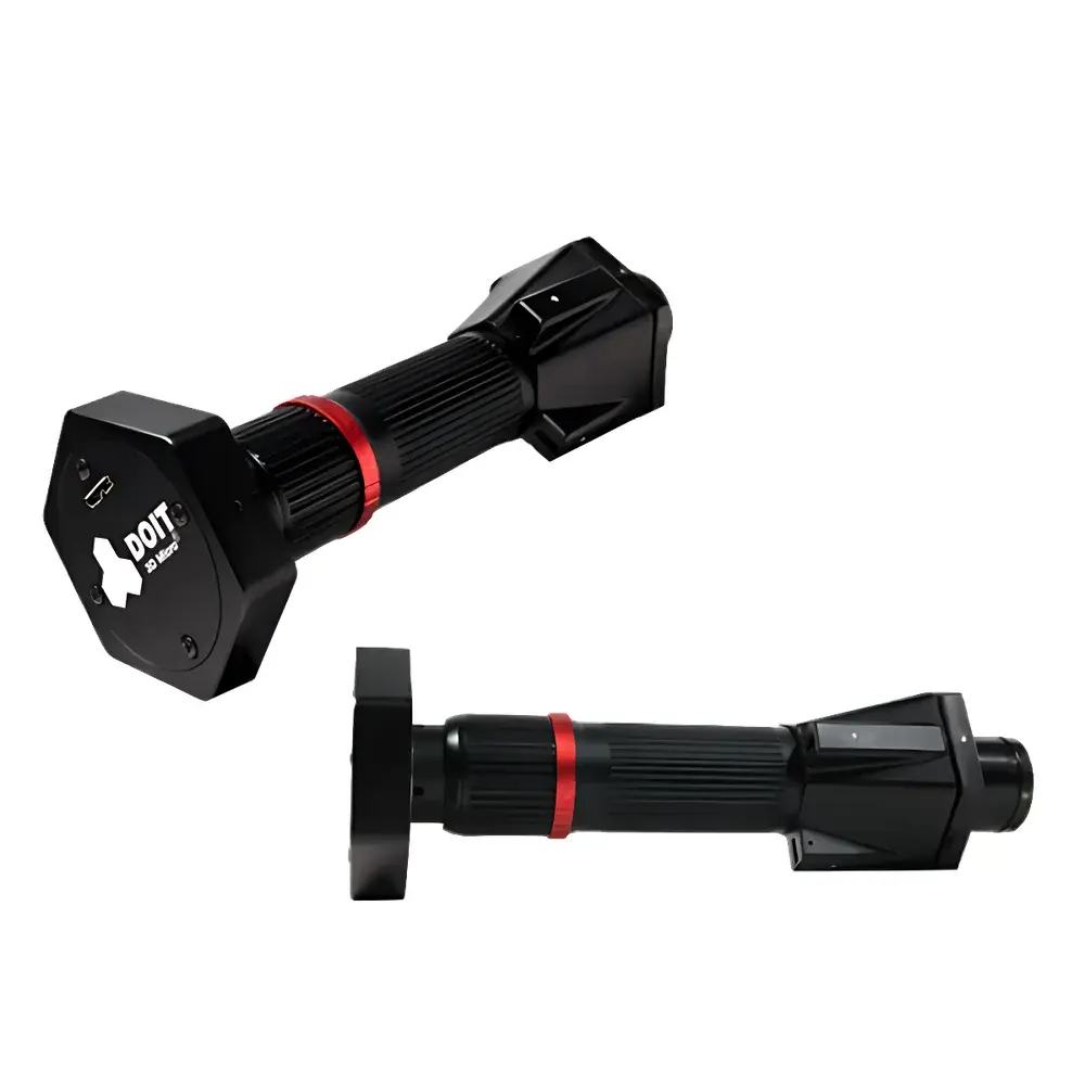
The Auniontech DOIT Fourier Light-Field Microscopy Camera Module is an engineered optical add-on system that transforms standard 2D optical microscopes into high-fidelity 3D digital imaging platforms without modifying the host microscope’s optical path. Based on Fourier-domain light-field imaging—also known as integral imaging—the module captures angular and spatial information simultaneously via a microlens array placed at the intermediate image plane (e.g., eyepiece port or C-mount camera adapter). This architecture enables single-shot acquisition of multi-perspective light-field data, which is computationally decoded to reconstruct volumetric information, including depth-resolved focal stacks, synthetic refocusing, and perspective-shifted views. Unlike conventional z-stack acquisition requiring mechanical stage movement, the DOIT module delivers real-time 3D visualization with no moving parts, making it suitable for dynamic biological specimens, live-cell observation, and time-sensitive industrial inspection tasks where axial scanning introduces motion artifacts or throughput limitations.
Key Features
- Plug-and-play integration compatible with any upright or inverted microscope equipped with standard eyepiece tubes (23.2 mm or 30 mm) or C-mount camera ports
- Single-exposure 3D acquisition enabling real-time volumetric rendering at native frame rates (up to 60 fps depending on host camera and processing configuration)
- Computational refocusing across continuous axial planes without physical lens adjustment
- Depth-encoded visualization via color-mapped topographic projection and quantitative depth maps
- Support for stereo-pair generation and VR-compatible stereoscopic output using virtual viewpoint synthesis
- Modular firmware architecture allowing application-specific calibration profiles for brightfield, fluorescence, phase contrast, and differential interference contrast (DIC) modalities
Sample Compatibility & Compliance
The DOIT module supports a broad range of specimen types—from fixed tissue sections and cultured monolayers to live zebrafish embryos, organoids, and microelectronic wafers—without requiring specialized sample preparation or embedding. Its non-invasive acquisition protocol aligns with Good Laboratory Practice (GLP) and ISO/IEC 17025-compliant workflows where minimal perturbation and traceable metadata are required. When integrated with FDA 21 CFR Part 11–compliant software environments, the system supports audit-trail logging of reconstruction parameters, user authentication, and electronic signatures for regulated life science applications. Calibration routines follow traceable NIST-referenced procedures for lateral resolution verification and axial step validation.
Software & Data Management
The proprietary DOIT Acquisition & Reconstruction Suite (v4.x) provides a modular GUI for real-time preview, post-acquisition volumetric processing, and export in standardized formats including TIFF (OME-TIFF), HDF5, and STL (for surface mesh export). Core algorithms include Wiener deconvolution-based light-field inversion, epipolar geometry correction, and GPU-accelerated ray tracing for synthetic aperture rendering. The software supports batch processing pipelines compliant with FAIR data principles (Findable, Accessible, Interoperable, Reusable), enabling integration with laboratory information management systems (LIMS) and cloud-based analysis platforms such as OMERO or QuPath. All raw light-field datasets retain full angular-spatial metadata, ensuring reproducibility and facilitating third-party algorithm development under open API documentation.
Applications
- Life Sciences: Live 3D tracking of neuronal calcium dynamics; volumetric monitoring of spheroid growth and drug-induced morphological changes; subcellular organelle localization in intact tissues
- Industrial Metrology: Non-contact 3D profiling of solder joints, PCB traces, and MEMS device surfaces; defect classification in semiconductor packaging using depth-aware segmentation
- Forensics & Materials Science: Multi-angle reconstruction of toolmark impressions; depth-resolved analysis of layered paint chips or fiber cross-sections
- Agricultural Quality Control: In-situ 3D assessment of grain morphology, fungal hyphae penetration, and seed coat integrity
- Education & Training: Interactive 3D histology modules for medical and veterinary curricula; remote-access virtual microscopy labs supporting collaborative annotation and depth-aware annotation tools
FAQ
What microscope configurations are supported?
The DOIT module interfaces with standard DIN 23.2 mm eyepiece tubes and C-mount (1″) camera ports. It is compatible with both transmitted-light and epi-fluorescence systems using objectives ranging from 10× to 60× NA 0.9.
Does the system require custom objective lenses?
No. The module operates independently of objective design and does not alter magnification or optical conjugation; it functions as a field-port imaging adapter.
Can axial resolution be improved beyond the specified minimum step size?
Yes—axial sampling density is software-defined during reconstruction; higher oversampling increases computational load but improves depth interpolation fidelity.
Is fluorescence compatibility validated across common emission bands?
Yes. Optical coatings and microlens transmission profiles are optimized for 400–700 nm, covering DAPI, FITC, TRITC, and Cy5 spectral ranges without spectral crosstalk.
How is calibration maintained across different laboratories?
Each unit ships with a serialized calibration certificate referencing NIST-traceable USAF 1951 target measurements and includes automated in-situ drift compensation routines activated during startup.