Auniontech OQ PathScope & OQ VetScope Portable Optical Coherence Tomography Systems
| Brand | Auniontech |
|---|---|
| Model | OQ PathScope / OQ VetScope |
| Type | Spectral-Domain OCT (SD-OCT) |
| Light Source | Fiber-Coupled Superluminescent Diode (SLD), Center Wavelength: 840 nm ± 10 nm |
| Spectral Detection | High-Resolution CMOS Linear Array Spectrometer |
| Axial Resolution (in air) | ≤ 7 µm |
| Imaging Depth (in tissue) | ≥ 2.5 mm |
| Scan Speed | Up to 47,000 A-scans/s |
| Footprint | < 300 × 250 × 150 mm (OQ VetScope) |
| Interface | USB 3.0 |
| Software Platform | Windows-based OCT acquisition & visualization suite with real-time B-scan rendering, volume reconstruction, and export in DICOM/RAW/TIFF formats |
| Compliance | Designed for research use |
Overview
Optical Coherence Tomography (OCT) is a non-invasive, label-free interferometric imaging modality that enables micron-scale, cross-sectional visualization of scattering biological tissues in real time. Based on low-coherence interferometry, OCT measures the time delay and intensity of back-reflected or back-scattered light from internal microstructures—without requiring physical sectioning or contrast agents. The Auniontech OQ PathScope and OQ VetScope are compact, research-grade spectral-domain OCT (SD-OCT) systems engineered for reproducible, high-fidelity structural imaging in preclinical and translational laboratory environments. Unlike time-domain OCT, SD-OCT employs a broadband light source and a high-speed spectrometer to acquire full depth profiles (A-scans) simultaneously, delivering superior signal-to-noise ratio, faster acquisition rates, and improved phase stability. These systems operate at a center wavelength of 840 nm—a well-established optical window balancing penetration depth and resolution in soft tissue—and achieve axial resolution ≤ 7 µm in air (≈ 5 µm in tissue), with imaging depths exceeding 2.5 mm in scattering media such as cornea, retina, skin, and ex vivo biopsy specimens.
Key Features
- Cost-optimized architecture: Integrates fiber-coupled superluminescent diodes (SLDs) with stabilized output power and reduced intensity noise—minimizing motion artifacts and improving image homogeneity without reliance on costly swept-source lasers or high-end broadband sources.
- Robust spectral detection: Employs a high-pixel-count CMOS linear array spectrometer, delivering enhanced thermal and mechanical stability compared to traditional CCD-based spectrometers—reducing calibration drift and enabling consistent performance across ambient lab conditions.
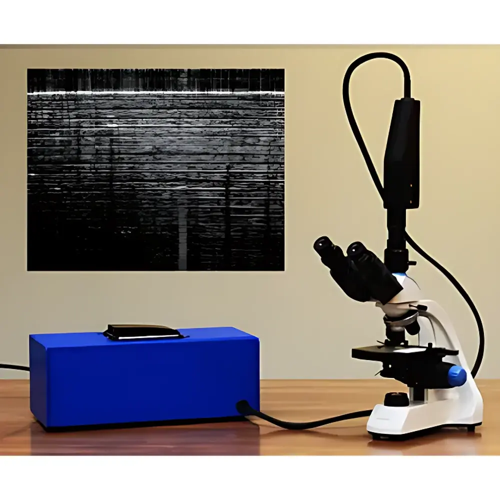
- Modular form factor: OQ VetScope (ultra-compact, benchtop-ready) and OQ PathScope (microscope-integrated variant) share identical core optics and data acquisition firmware, allowing seamless workflow scaling from rapid screening to high-magnification histopathological correlation.
- Real-time visualization: Onboard GPU-accelerated processing renders B-scans at up to 47,000 A-lines per second, with live depth-resolved preview, adjustable scan protocols (raster, radial, circular), and automatic dispersion compensation.
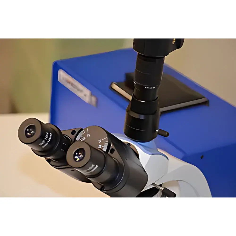
- Interoperable design: Standard C-mount and SM1-threaded optical interfaces enable direct coupling to upright or inverted research microscopes—including compatibility with 10×–40× objective lenses—for combined OCT-microscopy co-localization studies.
Sample Compatibility & Compliance
The OQ series supports diverse biological and biomaterial samples including excised ocular tissues (retina, sclera, cornea), rodent skin and cartilage, polymer phantoms, and layered hydrogel constructs. No fixation, staining, or dehydration is required. All systems are designated for in vitro and ex vivo research use only—not cleared for clinical diagnostic application. Manufacturing adheres to ISO 13485 principles for quality management of medical device-related R&D instrumentation. While not FDA 510(k)-cleared, the software architecture includes optional audit-trail logging, user access controls, and timestamped metadata embedding—facilitating alignment with Good Laboratory Practice (GLP) documentation standards and supporting traceability in regulated preclinical studies.
Software & Data Management
The bundled OCT Studio software provides a validated, Windows-based environment for system control, image acquisition, post-processing, and quantitative analysis. Core capabilities include automatic motion correction (via cross-correlation registration), logarithmic intensity scaling, speckle reduction filtering, layer segmentation (manual and semi-automated), and volumetric rendering (en face, 3D surface plots). Data exports conform to open scientific formats: raw interferogram matrices (.bin), calibrated intensity volumes (.tiff stack), and DICOM-compliant datasets (with modality tag “OT” for Optical Tomography). All acquisitions are automatically annotated with instrument serial number, date/time stamp, scan parameters, and user ID—ensuring full experimental provenance for peer-reviewed publication or internal reporting.
Applications
- Ophthalmic research: Retinal layer thickness mapping, optic nerve head topography, and glaucoma progression modeling in animal models.
- Dermatology & wound healing: Monitoring epidermal/dermal boundary dynamics, collagen remodeling, and vascular perfusion patterns in murine skin injury models.
- Pathology support: Rapid intraoperative margin assessment of excised tissue specimens prior to histology—reducing turnaround time and enabling targeted sectioning.
- Biomaterial characterization: Quantifying scaffold porosity, degradation kinetics, and cell infiltration depth in 3D-printed tissue engineering constructs.
- Neuroscience: Visualizing cortical lamination and myelin sheath integrity in fixed brain slices without sectioning-induced distortion.
FAQ
Is this system FDA-cleared for clinical use?
No. The OQ PathScope and OQ VetScope are intended exclusively for research use in laboratory settings and are not approved for human diagnostic applications.
Can I integrate this OCT system with my existing confocal or multiphoton microscope?
Yes—both models feature standardized optical interfaces (C-mount and SM1 threading) and provide mechanical and optical alignment guides for integration with commercial upright/inverted microscopes equipped with side ports or camera outputs.
What is the typical axial resolution in biological tissue?
Axial resolution is ≤ 7 µm in air and approximately 5 µm in tissue (e.g., retinal layers or dermis), depending on refractive index and scattering properties.
Does the software support batch processing of multiple OCT volumes?
Yes—OCT Studio includes scripting support (via Python API) for automated batch analysis, including layer thickness quantification, intensity profiling, and statistical comparison across experimental groups.
Is temperature stabilization required for stable long-term operation?
No active cooling is needed; the CMOS spectrometer and SLD source are designed for passive thermal management and exhibit minimal drift over 8-hour continuous operation within standard lab temperature ranges (18–25 °C).


