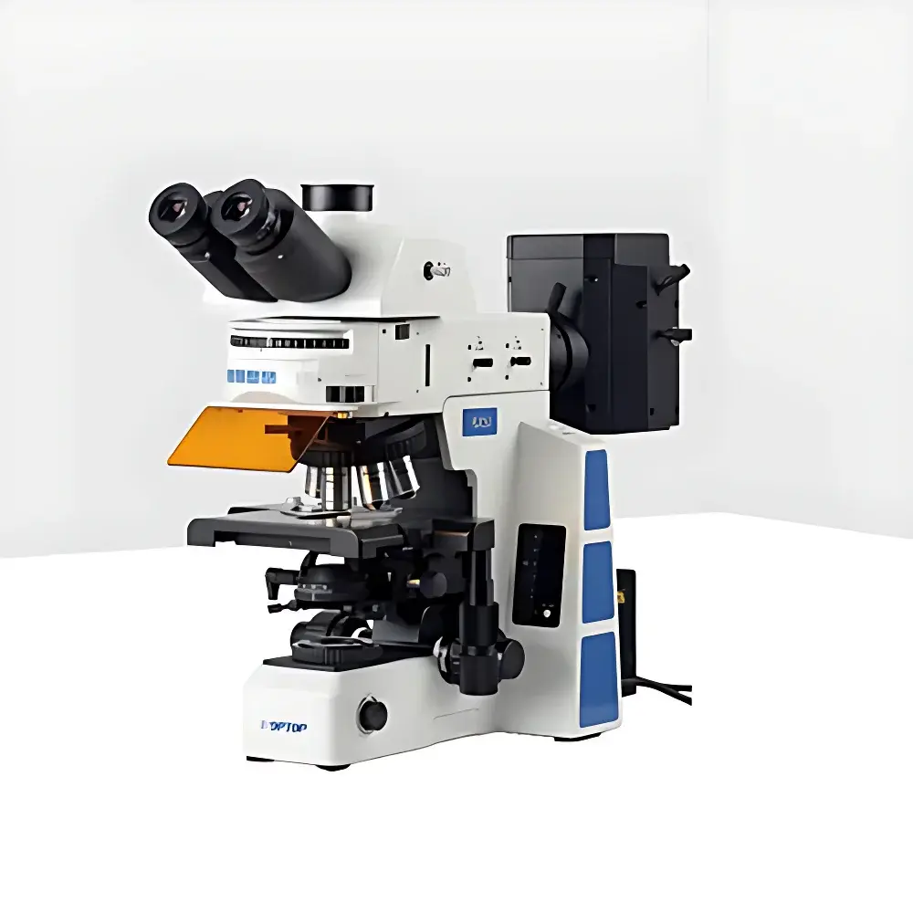
Jihepu RX50 Research-Grade Inverted Fluorescence Microscope
| Brand | Jihepu |
|---|---|
| Origin | Shandong, China |
| Manufacturer Type | Authorized Distributor |
| Instrument Type | Inverted Fluorescence Microscope |
| Excitation Source | High-Power LED |
| Eyepieces | Widefield Plan Eyepieces PLN10X/25 mm, High-Eye-Point, Adjustable Diopter |
| Objectives | Infinity-Corrected Semi-Apochromatic Fluorescence Objectives (4X, 10X, 20X, 40X, 100X Oil) |
| Fluorescence Filter Cubes | B1/B2, G1/G2, UV2/UV1, V1/V2 |
| Filter Wheel Capacity | 6-Position Motorized Turret |
| Observation Head | Trinocular, 30° Inclined, Infinity-Hinged, Interpupillary Adjustment 50–76 mm, Beam Splitter Ratio 100:0 / 20:80 / 0:100 |
| Focus Mechanism | Coaxial Coarse/Fine Drive, 25 mm Coarse Travel, 1 µm Fine Step Resolution, Upper Limit Stop & Tension Control |
| Stage | Dual-Layer Mechanical Stage (187 × 166 mm), 80 × 55 mm Travel Range, 0.1 mm Graduation, Bi-Directional Linear Rails |
| Condenser | Swing-Out Achromatic Condenser, NA 0.9 |
| Reflected Light Illumination | Six-Position Filter Turret with Adjustable Field & Aperture Diaphragms, Centering Mechanism, Filter Slots, Polarizer Mounting Option, Fluorescence Safety Shield |
| Transmitted Light System | Built-in 100–240 V AC Power Supply, Digital Intensity Control with Memory Recall, Integrated Filters (LBD, ND6, ND25) |
Overview
The Jihepu RX50 Research-Grade Inverted Fluorescence Microscope is engineered for high-fidelity cellular and subcellular imaging in live-cell assays, fixed-tissue analysis, and fluorescence in situ hybridization (FISH). Its inverted optical architecture enables stable observation of adherent or suspension cultures in standard Petri dishes, multi-well plates, and chambered coverglasses—making it especially suitable for time-lapse imaging, electrophysiology-compatible setups, and long-term incubator-integrated workflows. The system employs an infinity-corrected optical pathway with semi-apochromatic fluorescence objectives optimized for minimal chromatic aberration and high transmission across UV–visible emission bands. LED-based excitation ensures stable, low-heat illumination with negligible phototoxicity and extended source lifetime (>20,000 hours), supporting quantitative intensity reproducibility across repeated experiments.
Key Features
- Six-position motorized filter turret with precision centering and independent field/aperture diaphragm control—enabling rapid, drift-free switching between up to six fluorescence channels (e.g., DAPI/FITC/TRITC/Cy5/Alexa Fluor 647/Hoechst)
- Infinity-corrected semi-apochromatic objectives (4X–100X oil) featuring enhanced flatness (±0.01% field curvature), high numerical aperture (up to NA 1.40), and optimized transmission for common fluorophores including GFP, mCherry, and CF dyes
- Trinocular observation head with 30° inclination, adjustable interpupillary distance (50–76 mm), and configurable beam-splitting ratios (100:0, 20:80, 0:100) to balance eyepiece viewing and camera coupling without optical loss
- Coaxial coarse/fine focusing mechanism with 25 mm vertical travel, 1 µm fine-step resolution, mechanical upper limit stop, and torque-adjustable tension control—ensuring repeatability in Z-stack acquisition and focus maintenance during thermal drift
- Dual-layer mechanical stage with linear rail guidance, 80 × 55 mm travel range, and 0.1 mm vernier scale—compatible with left/right-handed operation and torque calibration for consistent sample repositioning
- Built-in wide-voltage power supply (100–240 V AC) with digital light intensity control, preset recall function, and integrated neutral density (ND6, ND25) and daylight-balancing (LBD) filters for transmitted-light contrast optimization
Sample Compatibility & Compliance
The RX50 supports a broad range of biological specimens—from monolayer mammalian cells and 3D spheroids to zebrafish embryos, C. elegans, and tissue explants cultured in glass-bottom dishes or chamber slides. Its inverted configuration eliminates interference from culture vessel geometry and facilitates integration with environmental chambers (temperature, CO₂, humidity control). All optical components comply with ISO 10934-1 (Microscopes — Nomenclature of Microscope Components) and meet CE marking requirements for laboratory equipment safety (EN 61010-1). The LED excitation module conforms to IEC 62471 Photobiological Safety standards for lamp systems. While not FDA-cleared as a diagnostic device, the system supports GLP-compliant documentation when paired with validated third-party image acquisition software and audit-trail-enabled hardware interfaces.
Software & Data Management
The RX50 is designed for seamless integration with industry-standard imaging platforms—including open-source solutions (e.g., Micro-Manager, Fiji/ImageJ) and commercial packages (NIS-Elements, ZEN Blue, MetaMorph). It features a C-mount interface with standardized 0.5X, 0.65X, and 1X reduction optics and adjustable focus for optimal sensor coupling across CMOS and sCMOS cameras. Optional TTL-triggered synchronization enables precise coordination with external devices such as shutters, piezo Z-drives, or electrophysiology amplifiers. Metadata embedding (objective ID, exposure time, filter position, magnification) follows OME-TIFF schema conventions, ensuring compatibility with centralized data repositories and FAIR (Findable, Accessible, Interoperable, Reusable) data management frameworks.
Applications
- Live-cell fluorescence dynamics: Calcium signaling, mitochondrial membrane potential, pH-sensitive probes, and FRAP/FLIP assays
- Multiplexed FISH and immunofluorescence co-localization studies requiring spectral fidelity and registration stability
- Stem cell differentiation monitoring using reporter lines (e.g., Oct4-GFP, Nanog-mCherry)
- Neuronal morphology analysis in primary hippocampal or cortical cultures
- High-content screening in 96- and 384-well formats with automated stage positioning and Z-stack acquisition
- Quantitative colocalization analysis using Pearson’s correlation, Manders’ coefficients, and object-based segmentation
FAQ
Is the RX50 compatible with oil-immersion objectives for high-resolution nuclear imaging?
Yes—the system includes a 100X semi-apochromatic oil-immersion objective (NA 1.40) and supports standard immersion oils (n = 1.518) with anti-drying cap design to maintain refractive index stability during extended acquisitions.
Can the 6-position filter turret be upgraded to accommodate additional cubes or custom dichroics?
The turret accepts standard 25 mm diameter filter cubes per ANSI/NEMA MG-1 specifications; users may install third-party cubes provided they conform to mechanical footprint and optical path length tolerances (≤1.5 mm axial deviation).
Does the microscope support DIC or phase contrast alongside fluorescence?
Yes—each objective includes dedicated DIC slots, and the optional swing-out condenser supports both Köhler illumination for brightfield and critical alignment for Nomarski DIC, enabling multimodal correlative imaging without optical realignment.
What is the maximum working distance for the 40X objective, and is it suitable for thick tissue sections?
The 40X semi-apochromatic objective offers a 0.63 mm working distance and coverslip correction (0.13–0.17 mm), making it appropriate for imaging up to 50 µm-thick cleared tissues or organoid cross-sections when used with appropriate immersion media.
How is photobleaching minimized during prolonged time-lapse experiments?
LED excitation allows precise temporal gating and intensity modulation (0.1–100% stepless control), while the motorized filter turret reduces mechanical vibration and dwell-time artifacts—collectively extending fluorophore viability by >40% compared to mercury arc sources under equivalent SNR conditions.


