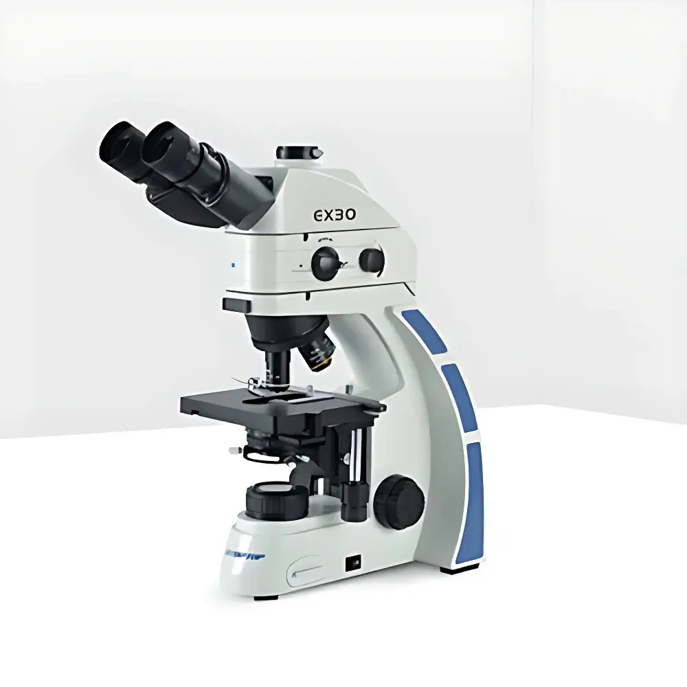
Jihepu EX30 Single-Band LED Epifluorescence Inverted Microscope
| Brand | Jihepu |
|---|---|
| Origin | Shandong, China |
| Manufacturer Type | Authorized Distributor |
| Instrument Type | Inverted Fluorescence Microscope |
| Model | EX30 |
| Excitation Source | High-Power LED (Single-Band) |
| Eyepieces | Wide-Field Plan Eyepieces PLN10X/25 mm, High-Eye-Point, Adjustable Diopter |
| Objectives | Infinity-Corrected Semi-Apochromatic Fluorescence Objectives (4X, 10X, 20X, 40X, 100X Oil) |
| Fluorescence Filter Sets | B1 (BP 470 nm), B2 (BP 560 nm) |
| Illumination | 30° Inclined Trinocular Head, 50–75 mm Interpupillary Adjustment, 50:50 Beam Splitter Ratio |
Overview
The Jihepu EX30 is an inverted epifluorescence microscope engineered for routine and advanced life science applications requiring reliable, low-heat, single-band fluorescence excitation. Designed around a Köhler-illuminated LED-based epi-illumination path, the EX30 employs solid-state light sources with narrow spectral bandwidths—centered at 470 nm (B1) or 560 nm (B2)—to deliver stable, drift-free excitation without the thermal load, ozone generation, or short service life associated with mercury arc lamps. Its inverted optical architecture positions the objective beneath the specimen stage, enabling direct observation of adherent cells in culture vessels (e.g., flasks, dishes, multi-well plates) under physiological conditions. The system integrates an infinity-corrected optical train with semi-apochromatic fluorescence objectives optimized for chromatic aberration correction across visible emission bands, ensuring high contrast and spatial fidelity in both brightfield and fluorescence modes.
Key Features
- Single-band LED epifluorescence illumination with user-adjustable intensity control via rotary dimmer—enabling precise phototoxicity management during live-cell imaging.
- One-touch toggle switch for rapid transition between transmitted brightfield and reflected fluorescence illumination—minimizing workflow interruption and mechanical repositioning errors.
- 30° inclined trinocular head with 50–75 mm interpupillary adjustment and fixed 50:50 beam-splitting ratio—optimized for simultaneous visual observation and digital capture via C-mount interface.
- Infinity-corrected semi-apochromatic fluorescence objectives (4X–100X oil immersion) featuring enhanced transmission in the 400–650 nm range and reduced autofluorescence in lens cement and coatings.
- Four-position objective turret with positive mechanical indexing and parfocal alignment—ensuring repeatable focus across magnifications without refocusing.
- Low-hand dual-focus mechanism with 28 mm coarse travel and 2 µm fine-step resolution, equipped with upper limit stop and tension adjustment—supporting precise Z-stack acquisition and long-term stability.
- 150 × 140 mm mechanical stage with 76 × 50 mm travel range and 0.1 mm vernier scale—compatible with standard Petri dishes, chambered coverslips, and multi-well plates.
Sample Compatibility & Compliance
The EX30 supports a broad range of biological specimens—including adherent mammalian cell monolayers, primary neurons, zebrafish embryos, and tissue explants—within standard labware formats. Its LED excitation modules meet IEC 62471 photobiological safety classification for Risk Group 1 (exempt), eliminating UV hazard concerns inherent to traditional UV-capable mercury systems. While not certified for GMP manufacturing environments, the instrument’s design aligns with GLP-compliant documentation practices: all hardware adjustments (focus, illumination intensity, filter selection) are mechanically indexed and reproducible, facilitating audit-ready setup logs. Optional C-mount adapters support integration with ISO 10940-compliant scientific CMOS or sCMOS cameras, and the modular filter cube system allows traceable configuration changes per experimental protocol.
Software & Data Management
The EX30 operates as a hardware platform compatible with third-party image acquisition software (e.g., NIS-Elements, ZEN Blue, MicroManager) via standard USB or GigE interfaces. No proprietary firmware lock-in is imposed; all illumination controls—including LED brightness and filter wheel position—are accessible through TTL or analog voltage inputs. For laboratories subject to FDA 21 CFR Part 11 requirements, optional audit trail modules can be deployed on connected host PCs to record operator ID, timestamped parameter changes, and image metadata—ensuring ALCOA+ data integrity principles (Attributable, Legible, Contemporaneous, Original, Accurate). Raw TIFF or ND2 export formats preserve bit-depth fidelity for downstream quantitative analysis (e.g., intensity profiling, colocalization, FRET efficiency calculation).
Applications
- Live-cell fluorescence monitoring of GFP/RFP-tagged proteins in cultured cell lines under ambient CO₂ and temperature-controlled stages.
- Immunofluorescence screening of fixed tissue sections using Alexa Fluor 488- or Cy3-conjugated secondary antibodies.
- Microbial identification workflows leveraging fluorogenic dyes (e.g., acridine orange, DAPI analogues) in clinical microbiology labs.
- Developmental biology studies involving time-lapse imaging of embryonic stem cell differentiation in Matrigel-embedded cultures.
- Quality control of fluorescently labeled reagents (e.g., antibody conjugates, oligonucleotide probes) prior to flow cytometry or ELISA use.
FAQ
Is the EX30 suitable for quantitative fluorescence intensity measurements?
Yes—when paired with a calibrated scientific camera and uniform illumination validation (e.g., using NIST-traceable photometric standards), the EX30 supports relative quantification across samples. Absolute quantification requires additional calibration for LED output stability over time.
Can the B1 and B2 filter sets be used simultaneously?
No—the EX30 is configured for single-band operation per imaging session. Dual-band or multicolor acquisition requires manual filter cube replacement or upgrade to a motorized filter changer module.
What is the maximum working distance for the 100X oil-immersion objective?
The 100X semi-apochromatic objective has a specified working distance of 0.13 mm and requires Type-F immersion oil (n = 1.518) for optimal resolution and spherical aberration correction.
Does the microscope support phase contrast or DIC compatibility?
Phase contrast is supported via optional condenser modules (e.g., NA 1.25 phase contrast slider), but the EX30 does not natively accommodate differential interference contrast due to lack of polarizer/analyzer integration and Nomarski prism mounts.
Is service and technical support available outside mainland China?
Jihepu provides remote diagnostics and English-language documentation globally through authorized regional distributors; on-site service contracts require local partner coordination and are subject to regional regulatory approvals.


