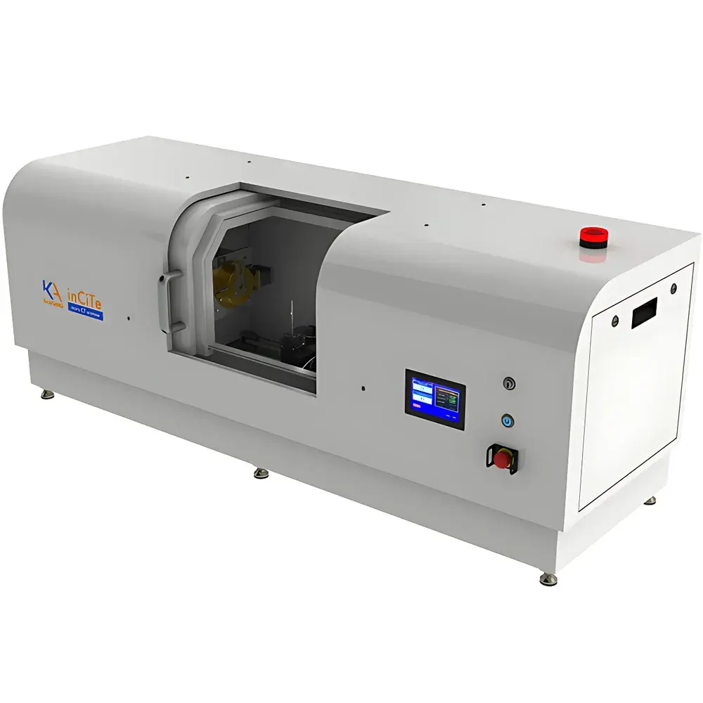
KA Imaging inCiTe CT Desktop X-ray Phase-Contrast Micro-CT System
| Brand | KA Imaging |
|---|---|
| Origin | Canada |
| Model | inCiTe CT |
| Detector | BrillianSe™ amorphous selenium (a-Se) |
| Imaging Principle | Propagation-based phase-contrast tomography |
| Spatial Resolution | Sub-micron to ~1 µm (system-dependent) |
| Field of View | Up to 100 mm diameter |
| Sample Max Height | 150 mm |
| X-ray Source | Microfocus tungsten-target tube (high-energy capable) |
| Energy Range | 30–90 kVp |
| Scan Time | Seconds to minutes per projection (depending on dose and resolution) |
| Compliance | CE-marked, ISO 13485-aligned design, GLP-ready data traceability |
Overview
The KA Imaging inCiTe CT is a research-grade, desktop-scale X-ray phase-contrast micro-computed tomography (micro-CT) system engineered for high-fidelity 3D non-destructive imaging of low-atomic-number (low-Z) materials. Unlike conventional absorption-based X-ray imaging—which relies on differential X-ray attenuation and yields poor contrast for soft tissues, polymers, composites, or porous biomaterials—the inCiTe CT leverages propagation-based phase-contrast imaging (PCI). This technique exploits the refraction of X-rays as they pass through interfaces with differing refractive indices, converting subtle phase shifts into measurable intensity variations at the detector plane. No optical elements, gratings, or synchrotron radiation are required. The system operates with a high-stability microfocus X-ray source and KA Imaging’s proprietary BrillianSe™ amorphous selenium (a-Se) flat-panel detector, enabling high detective quantum efficiency (DQE), excellent spatial resolution (~1 µm effective pixel pitch), and rapid acquisition—making it uniquely suited for laboratory-based quantitative morphology studies where synchrotron access is impractical.
Key Features
- Propagation-based phase-contrast imaging (PBI) implemented via free-space propagation—no optics, no gratings, no synchrotron dependency
- BrillianSe™ a-Se detector: high-resolution, high-DQE, low electronic noise, optimized for low-dose phase-sensitive acquisition
- Desktop footprint (< 1.2 m²) with full shielding; compliant with international radiation safety standards (IEC 61331-1)
- Integrated high-stability microfocus X-ray source (30–90 kVp), optimized for both high-energy penetration and phase sensitivity
- Motorized precision sample stage with 5-axis alignment (X/Y/Z/rotation/tilt), supporting large-volume scans up to 100 mm diameter × 150 mm height
- Real-time preview and iterative reconstruction pipeline supporting FDK, SART, and phase-retrieval algorithms (e.g., Paganin method)
Sample Compatibility & Compliance
The inCiTe CT is validated for imaging diverse low-Z and heterogeneous samples—including biological soft tissue (e.g., murine knee joints), porous bone scaffolds, polymer composites (e.g., Kevlar fiber laminates), lightweight concrete aggregates, additively manufactured metal parts with internal porosity, and clinical biopsy specimens. Its phase-contrast capability significantly enhances edge visibility and interfacial contrast without staining or sectioning. System design adheres to ISO 13485 principles for medical device-related R&D environments and supports audit-ready metadata logging aligned with GLP practices. Data export formats include DICOM, TIFF stack, and HDF5—ensuring compatibility with third-party analysis tools (e.g., Avizo, Dragonfly, ImageJ/Fiji). While not a Class II medical device, its architecture meets foundational requirements for FDA 21 CFR Part 11–compliant data integrity when deployed in regulated preclinical labs.
Software & Data Management
The inCiTe CT is operated via KA Imaging’s proprietary acquisition and reconstruction suite, featuring intuitive workflow-driven GUI, real-time projection monitoring, and automated beam-hardening correction. Reconstruction modules support both absorption-only and phase-enhanced volumetric rendering. Phase retrieval is implemented via single-distance Paganin-type algorithms with tunable regularization parameters. All scan parameters—including source settings, geometry calibration, detector gain maps, and reconstruction kernels—are embedded in image headers (DICOM SR or JSON sidecar files). Raw projections and reconstructed volumes are timestamped, user-tagged, and stored with SHA-256 checksums—enabling full traceability for peer-reviewed publications or regulatory submissions. Batch processing and Python API access facilitate integration into automated QA/QC pipelines.
Applications
- Biomedical research: 3D visualization of trabecular bone architecture around orthopedic implants (e.g., Ti-6Al-4V), cartilage-bone interface mapping, vascular corrosion casting analysis
- Materials science: Quantification of fiber orientation/distribution in polymer composites, pore network analysis in foams and aerogels, defect detection in LPBF-printed AlSi10Mg parts
- Geoscience & agronomy: Root-soil interaction modeling, pore structure evolution in lightweight aggregate concrete, seed coat microstructure assessment
- Electronics & packaging: Non-destructive inspection of underfill voids, solder joint integrity, and flex circuit delamination
- Preclinical imaging: Ex vivo specimen radiography with native soft-tissue contrast—complementing histology without embedding artifacts
FAQ
How does phase-contrast imaging differ from conventional absorption CT?
Phase-contrast imaging detects X-ray wavefront distortion (refraction) rather than attenuation. It provides orders-of-magnitude higher contrast for low-Z materials where absorption contrast is inherently weak.
Is synchrotron radiation required to operate the inCiTe CT?
No. The system uses a laboratory-grade microfocus X-ray source and relies on free-space propagation—eliminating the need for coherence-enhancing optics or accelerator facilities.
What is the typical spatial resolution achievable?
Effective isotropic voxel size ranges from ~0.5 µm to 5 µm depending on magnification, source focal spot size, and detector binning—validated per ASTM E2737-21 for micro-CT resolution reporting.
Can the system perform time-resolved (4D) scans?
Yes—when combined with programmable stage motion and triggered acquisition, dynamic processes such as fluid infiltration or mechanical loading can be captured at sub-minute temporal resolution.
Does the software support quantitative morphometric analysis?
Yes. Volume-rendered datasets are exportable to standard platforms (e.g., BoneJ, CTAn, SimpleITK) for porosity, thickness, connectivity, and anisotropy quantification per ISO 11064 and ASTM E112 protocols.


