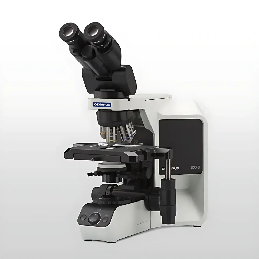
Olympus EVIDENT BX43 Manual Biological Microscope System
| Brand | Olympus/EVIDENT |
|---|---|
| Origin | Japan |
| Model | BX43 |
| Type | Modular Upright Biological Microscope for Brightfield, Phase Contrast, Polarization, and Fluorescence Imaging |
| Configuration | Manual Operation (No Motorized Components) |
| Optical Design | UIS2 Optical System |
| Compatibility | Standard 25 mm Field Number (FN25) Eyepieces & Accessories |
| Compliance | CE, ISO 9001, JIS B 7151 |
Overview
The Olympus EVIDENT BX43 is a high-precision, manually operated upright biological microscope system engineered for routine and advanced life science applications in academic laboratories, clinical pathology units, and quality control environments. Built upon Olympus’ proven UIS2 (Universal Infinity System) optical platform, the BX43 delivers consistent, high-fidelity image formation across multiple contrast modalities—including brightfield, phase contrast, polarization, and fluorescence—without optical compromise. Its rigid, thermally stable mechanical design minimizes stage drift and focus shift during extended observation sessions, supporting reproducible morphological assessment of fixed or live specimens. Unlike fully automated platforms, the BX43 prioritizes ergonomic manual operation, intuitive component interchangeability, and long-term serviceability—making it ideal for labs requiring reliability, regulatory traceability, and minimal software dependency.
Key Features
- Modular Architecture: Interchangeable optical and mechanical modules—including nosepieces (manual 4×–6×), observation tubes (binocular, trinocular, inclined/adjustable eyepiece angles), condensers (Abbe, AS, and LWD phase condensers), stages (mechanical XY with vernier scales, optional specimen holders), and intermediate optical units (e.g., fluorescence filter cubes, polarizing analyzers)—enable rapid reconfiguration for diverse imaging workflows.
- UIS2 Infinity-Corrected Optics: Delivers flat, high-resolution images across the full 25 mm field of view (FN25), with chromatic and spherical aberration correction optimized for both standard and high-NA objectives (up to 100× oil immersion).
- Ergonomic Design: Low-positioned focusing knobs, adjustable interpupillary distance, and tilt-compensated eyepiece tubes reduce operator fatigue during prolonged use—critical for histopathology screening and cell morphology training.
- Fluorescence Readiness: Pre-aligned light path supports standard fluorescence cube sets (e.g., FITC, TRITC, DAPI); optional mercury or LED illumination sources (e.g., U-HGLGPS or X-Cite series) integrate seamlessly via standardized dovetail mounts and power interfaces.
- Mechanical Stability: Cast-aluminum base and reinforced stand provide vibration damping; precision-ground stage rails ensure sub-micron positional repeatability over time—essential for comparative analysis and GLP-compliant documentation.
Sample Compatibility & Compliance
The BX43 accommodates standard glass slides (1 × 3 inches), petri dishes (up to 100 mm diameter with optional stage inserts), multi-well plates (6–96-well), and whole-mount tissue sections. It supports live-cell imaging when paired with environmental chambers (temperature/humidity/CO₂ control) and transmitted-light-compatible objectives. All optical components comply with ISO 10934-1 (microscope nomenclature), JIS B 7151 (Japanese industrial standard for microscopes), and CE marking requirements under EU Directive 2014/30/EU (EMC) and 2014/35/EU (LVD). The system meets essential requirements for use in ISO/IEC 17025-accredited testing laboratories and supports audit-ready documentation when integrated with validated digital imaging systems.
Software & Data Management
While the BX43 itself operates without embedded firmware or onboard software, its trinocular port and standardized C-mount interface (1× or 0.5× reduction) are fully compatible with third-party digital imaging platforms—including Olympus cellSens™ Desktop (v2.4+), ImageJ/Fiji, and HALCON-based custom acquisition software. When deployed in regulated environments, the system supports 21 CFR Part 11-compliant workflows via external image management servers that enforce user authentication, electronic signatures, and immutable audit trails for captured images and metadata (e.g., objective used, magnification, exposure settings, date/time stamp). No proprietary drivers or cloud dependencies are required for basic image capture or archiving.
Applications
- Routine histology and cytology screening in clinical pathology labs
- Teaching and demonstration of cellular structure, mitotic staging, and tissue architecture
- Quality assurance of cell culture monolayers and stem cell differentiation assays
- Material characterization of birefringent biological polymers (e.g., collagen, starch granules) using polarization optics
- Fluorescent localization studies with immunolabeled sections or transgenic reporter lines (e.g., GFP-tagged proteins)
- Pharmaceutical raw material identification per USP and EP 2.2.22 microscopy guidelines
FAQ
Is the BX43 compatible with motorized accessories or automated focus systems?
No—the BX43 is a purely manual platform. Motorized components (e.g., Z-drive, stage controllers, or filter turrets) are not supported natively and require upgrading to the BX53 or BX63 series.
Can I use third-party objectives with the BX43?
Yes, provided they conform to the UIS2 infinity-corrected standard (focal length 180 mm) and mechanical tube length specifications. Non-Olympus objectives may require individual parfocal adjustment and are not covered under warranty.
Does the BX43 meet FDA or ISO 13485 requirements for medical device manufacturing?
The BX43 itself is not a medical device but serves as a Class I support instrument. Its design and documentation support compliance with ISO 13485:2016 clause 7.6 (monitoring and measuring equipment) when calibrated and maintained per Olympus’ recommended service intervals.
What is the maximum working distance achievable with phase contrast on live samples?
Using the UPLSAPO 20× PH objective (WD = 0.6 mm) and an LWD PH condenser, the BX43 achieves up to 2.5 mm working distance with 40× PH objectives—sufficient for standard Petri dish and chamber slide configurations.


