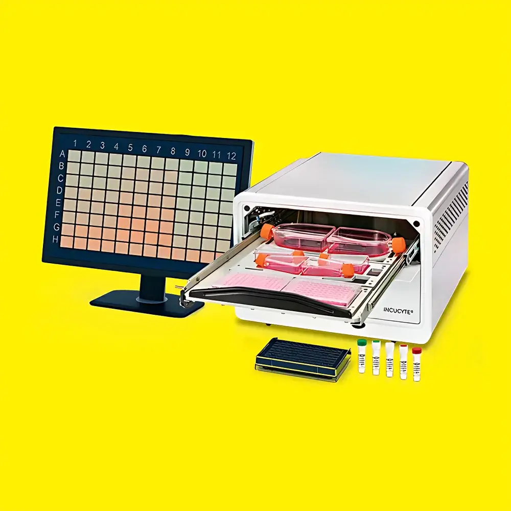
Sartorius Incucyte® S3 Live-Cell Analysis System
| Brand | Sartorius |
|---|---|
| Origin | USA |
| Manufacturer Type | Authorized Distributor |
| Origin Category | Imported Instrument |
| Model | Incucyte S3 |
| Pricing | Available Upon Request |
| Imaging Throughput | Experiment-Dependent |
Overview
The Sartorius Incucyte® S3 Live-Cell Analysis System is a fully integrated, incubator-based high-content imaging platform engineered for label-free and fluorescence-enabled longitudinal monitoring of living cells under physiologically relevant conditions. Utilizing automated, motorized optics and precision stage control, the system acquires high-definition (HD) phase contrast and dual-channel fluorescence (green and red) images directly inside standard CO₂ incubators—eliminating manual handling, environmental perturbation, and sampling bias. Its core measurement principle relies on quantitative image cytometry: time-lapse acquisition coupled with algorithm-driven segmentation, object classification, and kinetic parameter extraction (e.g., confluence, morphology metrics, fluorescent intensity kinetics, migration velocity, spheroid area/volume). Designed for reproducible, non-invasive observation across hours to months, the Incucyte S3 supports hypothesis-driven, dynamic phenotypic assays without endpoint lysis or fixation.
Key Features
- Incubator-integrated architecture: Mounts permanently inside standard 37 °C, 5% CO₂ tissue culture incubators; no external transfer required.
- Automated multi-well imaging: Supports simultaneous acquisition from up to six 6–384-well microplates per run, with independent acquisition schedules per plate.
- Optical stability design: Motorized objective turret and Z-stage maintain fixed focal plane during acquisition; optical components move—not the sample—ensuring minimal mechanical stress on adherent or suspension cultures.
- Dual-mode imaging: HD phase contrast (0.7×–20× magnification range) + two fluorescence channels (Ex/Em optimized for GFP/mCherry, FITC/TRITC, or custom dyes).
- Cloud-connected software: Browser-accessible interface with unlimited free user licenses; real-time remote monitoring and collaborative data review enabled via secure HTTPS.
- Zero-touch workflow: Guided experimental setup wizard, auto-calibration routines, and embedded metadata tagging ensure full traceability from protocol definition through analysis export.
Sample Compatibility & Compliance
The Incucyte S3 accommodates standard cell culture formats including flat-bottom and U-bottom 96-/384-well plates, chamber slides, Petri dishes (up to 100 mm), and specialized 3D scaffolds or organoid inserts. It is compatible with all common fluorophores, lentiviral reporters, CRISPR-edited biosensors, and commercially available Incucyte® reagents (e.g., Annexin V, Caspase-3/7, pHrodo™, Nuclight™). The system meets requirements for GLP-compliant workflows through audit-trail logging, electronic signature support (21 CFR Part 11-ready configuration), and version-controlled assay templates. Data files conform to open standards (TIFF, JSON, CSV) and are interoperable with third-party analysis platforms including ImageJ/Fiji, MATLAB, and Python-based scikit-image pipelines.
Software & Data Management
The Incucyte® Software v2023.x provides end-to-end analysis—from raw image ingestion to publication-ready visualization. Built-in algorithms perform adaptive thresholding, subcellular feature detection (e.g., neurite outgrowth, phagocytic cup formation), and kinetic modeling (sigmoidal growth, exponential decay, biphasic response). All processing steps are scriptable and repeatable; analysis parameters are saved with project metadata and can be reapplied across datasets. Export options include animated GIFs, multipanel time-series plots (with error bars), heatmaps, and annotated TIFF stacks. Raw and processed data are stored in hierarchical folder structures compliant with FAIR principles (Findable, Accessible, Interoperable, Reusable); optional integration with LIMS and ELN systems is supported via RESTful API.
Applications
The Incucyte S3 enables quantitative longitudinal assessment across diverse biological contexts: real-time cytotoxicity screening (IC₅₀ determination), immune-oncology assays (CAR-T or NK-cell mediated tumor spheroid killing), wound healing and chemotaxis quantification, stem cell differentiation tracking, neurosphere expansion dynamics, viral cytopathic effect monitoring, and autophagy flux analysis. Its validated use spans academic research, biopharma R&D (including IND-enabling studies), and CMC process development. Over 4,000 peer-reviewed publications—indexed in PubMed and cited in journals such as Nature Communications, Cell Reports, and Science Translational Medicine—demonstrate methodological rigor and cross-laboratory reproducibility.
FAQ
Can the Incucyte S3 be used outside a CO₂ incubator?
No—the system is designed exclusively for integration within standard humidified CO₂ incubators (37 °C, 5% CO₂); environmental control is maintained solely by the host incubator.
Does it support time-lapse imaging at sub-minute intervals?
Yes—acquisition frequency is user-defined down to 1-minute intervals per well, constrained only by total experiment duration and storage capacity.
Is offline analysis possible after data acquisition?
Yes—full analysis functionality remains available without network connectivity; cloud sync occurs only upon user-initiated upload or scheduled backup.
Are custom fluorescence filters supported?
The system ships with fixed GFP/mCherry filter sets; third-party filter cubes may be installed by qualified field service engineers following optical recalibration.
How is data integrity ensured during long-term experiments?
Continuous checksum validation, redundant local storage (RAID 1), and automatic recovery from transient power loss ensure zero frame loss over multi-week acquisitions.


