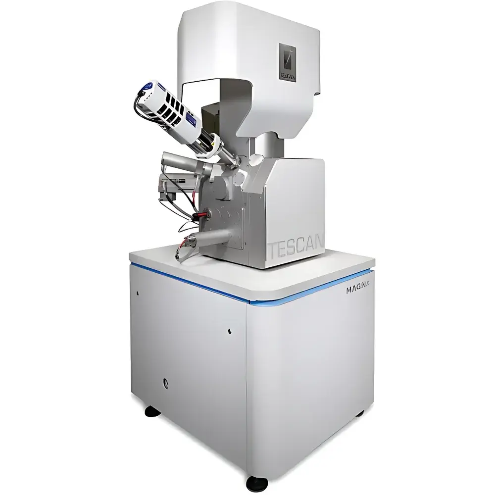
TESCAN MAGNA Ultra-High-Resolution Field-Emission Scanning Electron Microscope
| Brand | TESCAN |
|---|---|
| Origin | Czech Republic |
| Model | TESCAN MAGNA |
| Instrument Type | Floor-Standing SEM |
| Electron Gun | Schottky Field-Emission |
| SEM Class | Ultra-High-Resolution FE-SEM |
| Secondary Electron Resolution | 0.6 nm @ 15 kV |
| Magnification Range | 7× to 2,000,000× |
| Accelerating Voltage | 200 V – 30 kV (down to 50 V in deceleration mode) |
| Backscattered Electron Resolution | 1.6 nm @ 15 kV |
Overview
The TESCAN MAGNA is an ultra-high-resolution field-emission scanning electron microscope (FE-SEM) engineered for nanoscale surface characterization, microstructural analysis, and advanced analytical applications in materials science, semiconductor R&D, life sciences, and nanotechnology. Built around the proprietary Triglav™ SEM column—a tri-lens optical architecture—the MAGNA delivers exceptional resolution, signal fidelity, and operational stability across a wide voltage range (50 V–30 kV). Its core measurement principle relies on focused electron beam–sample interactions, generating secondary electrons (SE), backscattered electrons (BSE), and characteristic X-rays for topographic, compositional, and crystallographic analysis. The system’s low-voltage performance—enabled by optimized beam deceleration, high-efficiency in-column detection, and energy-filtered signal acquisition—makes it uniquely suited for beam-sensitive, non-conductive, and uncoated specimens without compromising spatial resolution or surface sensitivity.
Key Features
- Triglav™ Tri-Lens Column Architecture: Integrates three independently optimized objective lenses—UH-resolution lens for sub-nanometer imaging, Analytical lens with zero magnetic field at the sample for EDS/EBSD of magnetic materials, and Multi-Mode lens for dynamic beam shaping and multi-signal optimization.
- TriBE™ BSE Detection System: Three angle- and energy-selective backscattered electron detectors: in-column f-BSE (axial), mid-angle BSE, and chamber-mounted wide-angle BSE—enabling atomic-number contrast, channeling contrast, and topographic differentiation in a single acquisition.
- TriSE™ SE Detection System: Three complementary secondary electron detectors—including in-column In-Beam SE (for ultra-short working distance), BDM-mode SE (optimized for high-resolution low-kV imaging), and chamber-mounted SE (for robust topographic contrast)—support simultaneous multi-signal imaging with independent gain control.
- Schottky Field-Emission Electron Source: Delivers stable, high-brightness emission with beam currents up to 400 nA and rapid voltage switching (<100 ms), ensuring consistent signal-to-noise ratio across analytical modes including EDS, WDS, and EBSD.
- Adaptive Beam Optimization: Real-time beam convergence and spot size correction under varying probe current and acceleration conditions, maintaining optimal resolution during long-duration mapping or large-area stitching.
Sample Compatibility & Compliance
The MAGNA supports diverse sample types without mandatory conductive coating—including insulating ceramics, hydrated biological tissues, polymer blends, photoresists, and delicate 2D materials—thanks to its low-voltage imaging capability (down to 50 V) and charge compensation via beam deceleration and gas injection (optional). The system complies with international standards for laboratory instrumentation, including ISO/IEC 17025 requirements for calibration traceability, CE marking per EU Machinery Directive 2006/42/EC, and electromagnetic compatibility (EMC) per EN 61326-1. For regulated environments, optional audit-trail-enabled software modules support 21 CFR Part 11 compliance, facilitating GLP/GMP-aligned workflows in pharmaceutical development and medical device characterization.
Software & Data Management
TESCAN Essence™ is a modular, multi-user software platform designed for reproducible, workflow-driven operation. Its Layout Manager enables user-defined interface configurations—tailored for routine QC, failure analysis, or research-grade tomography—with role-based access control and session logging. Automated acquisition wizards guide users through stage calibration, focus/stigmation routines, and EDS map setup. All acquired images, spectra, and metadata are stored in vendor-neutral formats (TIFF, HDF5, .emsa) with embedded EXIF-like tags (accelerating voltage, dwell time, detector settings, stage coordinates). Raw data export supports third-party processing tools (e.g., HyperSpy, DigitalMicrograph), while built-in batch processing enables automated particle analysis, grain sizing, and phase quantification using machine-learning-assisted segmentation models.
Applications
- Nanomaterial Characterization: High-fidelity imaging of nanoparticles, nanowires, MOFs, and graphene derivatives at <1 nm resolution—even at ≤1 kV—to preserve surface chemistry and minimize beam damage.
- Semiconductor Metrology: Critical dimension (CD) measurement, defect review, and cross-sectional analysis of FinFETs, GAA transistors, and advanced packaging interconnects using low-kV SE/BSE dual-channel imaging.
- Life Sciences: Uncoated cryo- or ambient-conditioned biological specimens (e.g., extracellular vesicles, collagen fibrils, neural tissue sections) imaged with minimal charging artifacts and enhanced surface contrast.
- Geosciences & Ceramics: Quantitative mineral phase mapping via integrated EBSD-EDS correlation, grain boundary analysis, and porosity assessment in sintered oxides and composite refractories.
- Failure Analysis: In situ electrical biasing (with optional probes) combined with real-time SE/BSE monitoring for dynamic observation of electromigration, dielectric breakdown, or thermal degradation mechanisms.
FAQ
What is the minimum accelerating voltage achievable in deceleration mode?
The MAGNA achieves a landing energy as low as 50 V using beam deceleration, enabling high-contrast imaging of ultra-sensitive samples without charge buildup.
Does the system support automated EBSD indexing and orientation mapping?
Yes—when equipped with a compatible EBSD detector and TESCAN’s Crystal™ software module, the MAGNA performs fully automated pattern indexing, phase identification, and grain reconstruction with sub-degree angular precision.
Can the TriSE™ detectors operate simultaneously during a single scan?
Yes—up to three SE detectors can be acquired in parallel with independent gain, offset, and dwell time settings, allowing real-time multi-contrast fusion and differential signal subtraction.
Is remote operation supported for multi-site laboratories?
Essence™ includes secure remote desktop access with TLS-encrypted session handshaking, enabling supervised operation, troubleshooting, and training from off-site locations while preserving local audit trails.
How is beam stability maintained during extended EDS mapping sessions?
The Triglav™ column incorporates active thermal drift compensation and real-time beam alignment feedback, reducing positional drift to <5 nm/hour—critical for high-resolution spectral imaging over multi-hour acquisitions.


