TESCAN CoreTOM / DynaTOM / UniTOM HR Industrial Micro-CT Systems
| Brand | TESCAN |
|---|---|
| Origin | Czech Republic |
| Model Series | CoreTOM, DynaTOM, UniTOM HR |
| Spatial Resolution (min) | 0.6 µm (UniTOM HR), 3 µm (CoreTOM & DynaTOM, JIMA mode) |
| Max Sample Dimensions | Ø300 mm × H500 mm (UniTOM HR), up to 1150 mm length (CoreTOM) |
| Scan Speed | Full tomogram in seconds (DynaTOM), high-throughput multi-scale acquisition (CoreTOM), configurable acquisition timing (UniTOM HR) |
| System Architecture | Fixed-sample geometry with rotating X-ray source/detector (DynaTOM) |
Overview
The TESCAN CoreTOM, DynaTOM, and UniTOM HR series constitute a comprehensive family of laboratory-based industrial micro-computed tomography (micro-CT) systems engineered for non-destructive 3D and 4D structural characterization across scientific and industrial domains. Each platform implements cone-beam X-ray computed tomography based on filtered back-projection and iterative reconstruction algorithms, delivering quantitative volumetric data with spatial resolution ranging from sub-micron (600 nm in UniTOM HR) to micrometer-scale (3 µm in CoreTOM and DynaTOM under JIMA resolution mode). Unlike synchrotron-based solutions, these systems operate with sealed-tube or open-tube microfocus X-ray sources (typically 20–225 kV), coupled with high-dynamic-range flat-panel detectors or photon-counting CMOS sensors. The core measurement principle relies on angularly resolved projection acquisition—either via sample rotation (CoreTOM, UniTOM HR) or source/detector rotation around a stationary specimen (DynaTOM)—followed by volumetric reconstruction using GPU-accelerated software pipelines compliant with ISO 12799:2021 (non-destructive testing — industrial computed tomography — vocabulary and definitions) and aligned with ASTM E1441-22 guidelines for CT system performance verification.
Key Features
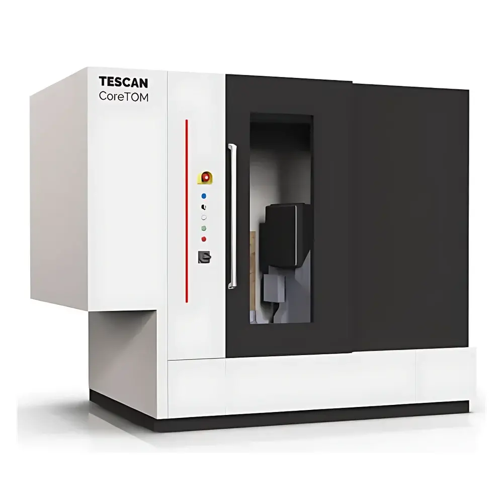
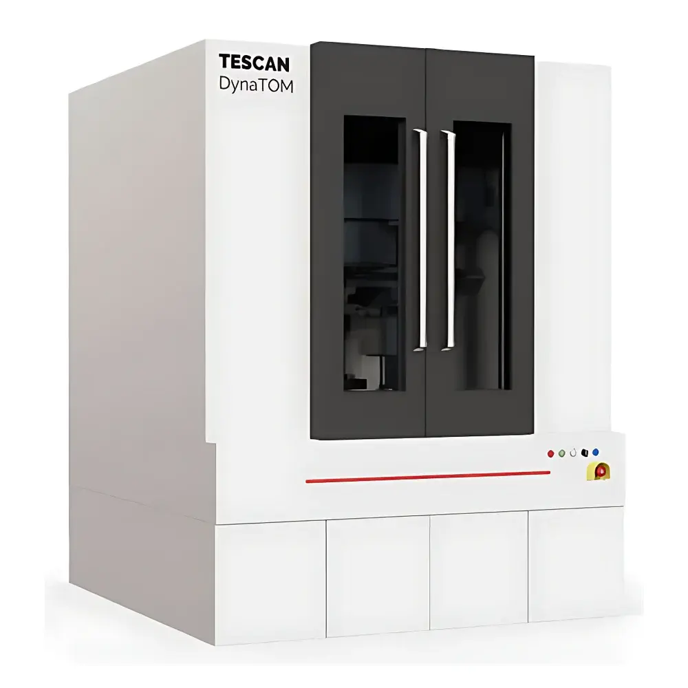
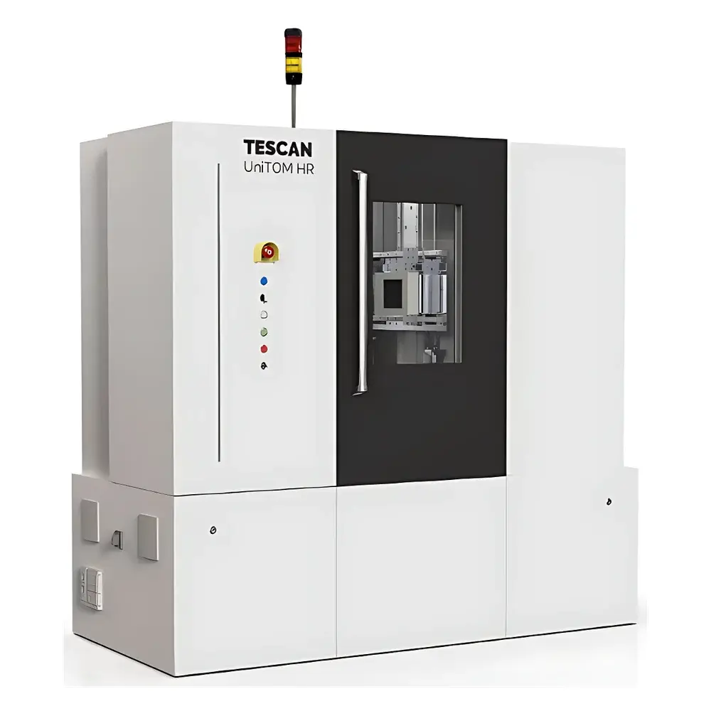
- Three purpose-built platforms: CoreTOM for multi-scale imaging of large-volume specimens (up to 1.15 m rock cores); DynaTOM for time-resolved in situ monitoring with sub-second full-tomogram acquisition; UniTOM HR for ultimate spatial fidelity (≤0.6 µm real resolution) and hardware modularity.
- DynaTOM’s fixed-sample architecture enables seamless integration with external stimuli: hydraulic pressure cells, electrochemical flow cells, thermal stages, and mechanical testers—all without compromising geometric stability or scan repeatability.
- CoreTOM employs adaptive field-of-view (FOV) navigation: users acquire low-resolution overview scans first, then define regions-of-interest (ROIs) for targeted high-resolution rescan—preserving global context while optimizing beamtime and data storage.
- UniTOM HR supports multi-source/multi-detector configurations, including dual-energy and phase-contrast modalities, with hardware-level synchronization for correlative 4D experiments (e.g., simultaneous mechanical loading + CT).
- All systems feature TESCAN’s proprietary Amira-Avizo-powered reconstruction and analysis suite, supporting automated pore network extraction, grain segmentation, defect quantification, and morphometric statistics per ISO/IEC 17025-accredited workflows.
Sample Compatibility & Compliance
The TESCAN micro-CT platforms accommodate heterogeneous samples spanning geological cores, additively manufactured metal parts, battery electrodes, composite laminates, biological tissues (fixed or stained), and porous ceramics. Maximum specimen dimensions range from Ø300 mm × H500 mm (UniTOM HR) to 1150 mm length (CoreTOM). Sample handling complies with ISO 15530-3:2020 (coordinate metrology — calibration of coordinate measuring machines — part 3: use of calibrated workpieces) for dimensional traceability. Data integrity adheres to FDA 21 CFR Part 11 requirements through audit-trail-enabled acquisition logs, electronic signatures, and version-controlled reconstruction parameters. System validation protocols align with ASTM E2865-22 (standard guide for validation of micro-CT systems) and support GLP/GMP-regulated environments where raw projection data, metadata, and processing history must be retained for regulatory review.
Software & Data Management
Acquisition, reconstruction, and analysis are unified within TESCAN’s integrated software environment, built on the Amira-Avizo platform with dedicated CT modules. Real-time preview, dose optimization, and automatic center-of-rotation correction are embedded at the acquisition layer. Reconstruction leverages both CPU-based FDK and GPU-accelerated SART/SIRT algorithms, with optional AI-assisted denoising (via trained convolutional neural networks) to enhance SNR without sacrificing resolution. All datasets are stored in DICOM-compliant format with embedded EXIF-like metadata (kV, µA, exposure time, detector binning, geometric magnification). Project archives include full provenance tracking—linking raw projections to reconstructed volumes, segmentation masks, and quantitative reports—for reproducibility and audit readiness. Export options cover STL, VTK, TIFF stacks, and HDF5 for integration into finite element analysis (FEA) or machine learning pipelines.
Applications
- Petroleum Geoscience: Quantitative pore-throat network modeling, fluid saturation dynamics, and diagenetic mineral mapping in whole-core samples under reservoir-relevant confining pressures.
- Advanced Manufacturing: Defect detection (porosity, lack-of-fusion, cracking) in AM Ti-6Al-4V and Inconel 718 components; lattice structure fidelity assessment; post-heat-treatment microstructural evolution.
- Energy Materials: In situ lithium-ion battery electrode degradation during cycling; solid-electrolyte interphase (SEI) growth kinetics; cathode particle fracture analysis.
- Materials Science: 4D creep and fatigue studies in metallic alloys; fiber orientation distribution in carbon-fiber composites; ice nucleation and dendritic growth in cryo-CT.
- Life Sciences: High-resolution vasculature mapping in decalcified bone; trabecular architecture quantification per ASBMR standards; scaffold porosity and interconnectivity in tissue engineering constructs.
FAQ
What is the difference between CoreTOM and UniTOM HR in terms of resolution and application focus?
CoreTOM prioritizes large-volume, multi-scale imaging (e.g., meter-scale rock cores) with resolution down to 3 µm; UniTOM HR delivers sub-micron resolution (≤0.6 µm) for small-to-medium specimens requiring maximum detail, such as battery particles or MEMS devices.
Can DynaTOM perform true 4D imaging (3D + time) with sub-second temporal resolution?
Yes—DynaTOM acquires complete tomographic volumes in ~1–5 seconds depending on voxel size and signal-to-noise requirements, enabling continuous acquisition over hours or days for dynamic process monitoring.
Is hardware upgrade support available for UniTOM HR after initial installation?
Yes—UniTOM HR uses a modular chassis design; additional X-ray sources, detectors, or motion stages can be integrated without system decommissioning, preserving long-term ROI and technical adaptability.
Do these systems comply with international standards for quantitative CT metrology?
All models support traceable calibration using NIST-traceable phantoms; reconstruction outputs meet ISO 12799 and ASTM E2865 requirements for dimensional accuracy, contrast-to-noise ratio (CNR), and spatial resolution reporting.
How is radiation safety managed during operation?
Each system is fully shielded per IEC 61000-6-4 and local regulatory codes (e.g., German RöV, US NRC 10 CFR 35); interlocked enclosures, real-time dose monitoring, and automated beam shutters ensure ALARA-compliant operation without external shielding infrastructure.

")

