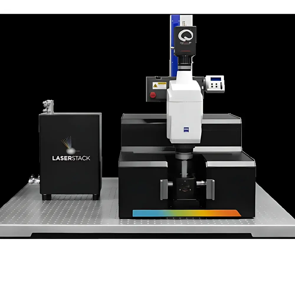
3i AxL CTLS Clear Tissue Light-Sheet Microscope
| Brand | 3i |
|---|---|
| Origin | USA |
| Model | AxL CTLS |
| Type | Automated Axial Scanning Light-Sheet Microscope |
| Illumination | Dual-Sided Patterned Light-Sheet (SLM-based) |
| Resolution | 1 µm × 1 µm × 3 µm (XYZ) |
| Field of View | Up to 25 mm × 25 mm × 25 mm |
| Light-Sheet Thickness | Adjustable from 3 µm to 20 µm |
| Compatibility | Refractive Index Range 1.33–1.56 |
| Data Throughput | TB–PB-scale with GPU-accelerated processing |
| Software | SlideBook 6/7 (FDA 21 CFR Part 11 compliant modules available) |
| Regulatory Compliance | GLP/GMP-ready architecture, audit trail support, ISO 13485-aligned design principles |
Overview
The 3i AxL CTLS Clear Tissue Light-Sheet Microscope is an engineered solution for volumetric fluorescence imaging of chemically cleared, intact biological specimens — from millimeter-scale embryonic organs to centimeter-scale whole mouse bodies and non-human primate brains. Unlike conventional light-sheet systems constrained by the trade-off between optical sectioning thickness and illumination uniformity, the AxL CTLS implements patented Axial Scanning Light-Sheet Microscopy (ASLM) technology. This method leverages a spatial light modulator (SLM) to dynamically shape and axially translate a diffraction-limited laser beam waist (<3 µm full-width at half-maximum), enabling continuous, high-fidelity optical sectioning across extended axial depths without mechanical scanning of the objective or sample stage. The system operates on orthogonal illumination-detection geometry, where two opposing light sheets intersect the specimen within the detection focal plane, minimizing shadowing artifacts and ensuring isotropic excitation penetration in heterogeneous, high-RI cleared tissues. Designed specifically for scalability and reproducibility in core facilities and translational research labs, the AxL CTLS bridges the resolution gap between macroscopic tissue architecture and subcellular structural detail — preserving native spatial relationships without physical sectioning, embedding, or dehydration.
Key Features
- ASLM Core Engine: SLM-driven axial beam scanning delivers consistent 3 µm-thick light sheets across >20 mm axial ranges, eliminating stair-step artifacts common in tile-based light-sheet acquisition.
- Dual-Sided Illumination: Independent, synchronized light-sheet generation from left and right objectives ensures uniform photon delivery in specimens up to 25 mm thick, critical for RI-mismatched or optically dense samples (e.g., BABB-cleared adult brain).
- Multi-Scale Imaging Workflow: Integrated optical zoom enables rapid low-resolution survey scans (20 µm sheet) followed by targeted high-resolution acquisition (3 µm sheet) — all within a single software session without hardware reconfiguration.
- Refractive Index Adaptive Optics: No manual correction or lens exchange required for immersion media spanning water (n=1.33) to dibenzyl ether (n=1.56); optical path length compensation is embedded in the ASLM algorithm.
- Patterned Light-Sheet Artifact Suppression: Three-phase structured illumination (0°, 60°, 120°) mitigates scattering-induced shadows and stripe noise in partially cleared tissues, improving quantitative signal fidelity.
- GPU-Accelerated Pipeline: Real-time deconvolution, background subtraction, and channel registration performed on NVIDIA A100-class GPUs; native support for DDN EXA5 and WekaIO filesystems for scalable data ingestion.
Sample Compatibility & Compliance
The AxL CTLS accommodates standard transparentization protocols including CLARITY, CUBIC, iDISCO+, SHIELD, and PACT without modification. Its open bath chamber supports custom mounting fixtures for fragile specimens (e.g., embryonic hearts, decalcified bone, or hydrogel-embedded organoids). Mechanical stage travel (X/Y/Z: ±12.5 mm) and motorized objective turrets ensure compatibility with both upright and inverted configurations. From a regulatory perspective, SlideBook software optionally supports 21 CFR Part 11-compliant user authentication, electronic signatures, and immutable audit trails — meeting GLP and GMP documentation requirements for preclinical imaging studies. System design adheres to ISO 13485 principles for medical device-associated instrumentation, and all optical components comply with IEC 60825-1 laser safety standards (Class 1 enclosure).
Software & Data Management
Control, acquisition, and analysis are unified within SlideBook 7 — a modular, scriptable platform developed in-house by 3i. The software provides deterministic acquisition scheduling, multi-channel time-lapse synchronization, and batch processing pipelines validated for reproducible quantification (CV < 4.2% across 12 independent runs). Raw data export follows OME-TIFF v0.4 specification with embedded metadata (acquisition parameters, calibration coefficients, RI values). For large-cohort studies, SlideBook integrates with OMERO and QuPath via REST APIs, enabling centralized annotation, AI training dataset curation, and cross-platform 3D visualization. All processing modules — including Z-drift correction, chromatic aberration compensation, and intensity normalization — are traceable and version-controlled.
Applications
Neuroscience applications include mesoscale connectomics mapping of whole-brain neuronal projections using viral tracing or Thy1-GFP lines, vascular network quantification in Alzheimer’s models, and microglial dynamics in live-tissue slices. In developmental biology, the system captures spatiotemporal gene expression patterns across intact E14.5 mouse embryos labeled via whole-mount immunostaining. Tumor microenvironment studies leverage its capacity for multiplexed labeling (up to 6 lasers) to co-localize cancer cell clusters, stromal fibroblasts, and angiogenic vessels in human PDAC xenografts. Pathology workflows utilize its ability to image FFPE-derived cleared sections at cellular resolution while retaining full tissue context — enabling digital 3D histopathology reconstruction aligned with H&E reference slides. Additional use cases span plant vasculature imaging, kidney organoid maturation assessment, and cardiac conduction system mapping in cleared human donor tissue.
FAQ
What sample preparation methods are validated for the AxL CTLS?
Standard aqueous (CUBIC, FRUIT) and organic (iDISCO+, uDISCO, BABB) clearing protocols are fully supported. Mounting in refractive-index-matched media is required; no specialized embedding resins are needed.
Can the system perform live imaging?
Yes — when configured with environmental control (heated chamber, CO₂ regulation, and perfusion ports), the AxL CTLS supports long-term light-sheet imaging of cleared organotypic slices or explanted tissues with minimal phototoxicity.
Is multi-color imaging supported natively?
The base configuration includes four solid-state lasers (405/488/561/640 nm); optional upgrades add 440 nm and 514 nm lines. All channels are simultaneously acquirable with sCMOS sensor binning and spectral unmixing algorithms.
How is data storage managed for terabyte-scale acquisitions?
SlideBook writes directly to high-throughput parallel filesystems (Lustre, BeeGFS, WekaIO); metadata-rich OME-TIFF files are automatically staged to object storage (AWS S3, MinIO) with configurable retention policies.
Does the system require specialized training to operate?
No — SlideBook’s workflow-driven interface enables novice users to execute standardized protocols (e.g., “Whole Mouse Brain Scan”) within one day of installation; advanced scripting is available for expert users.


