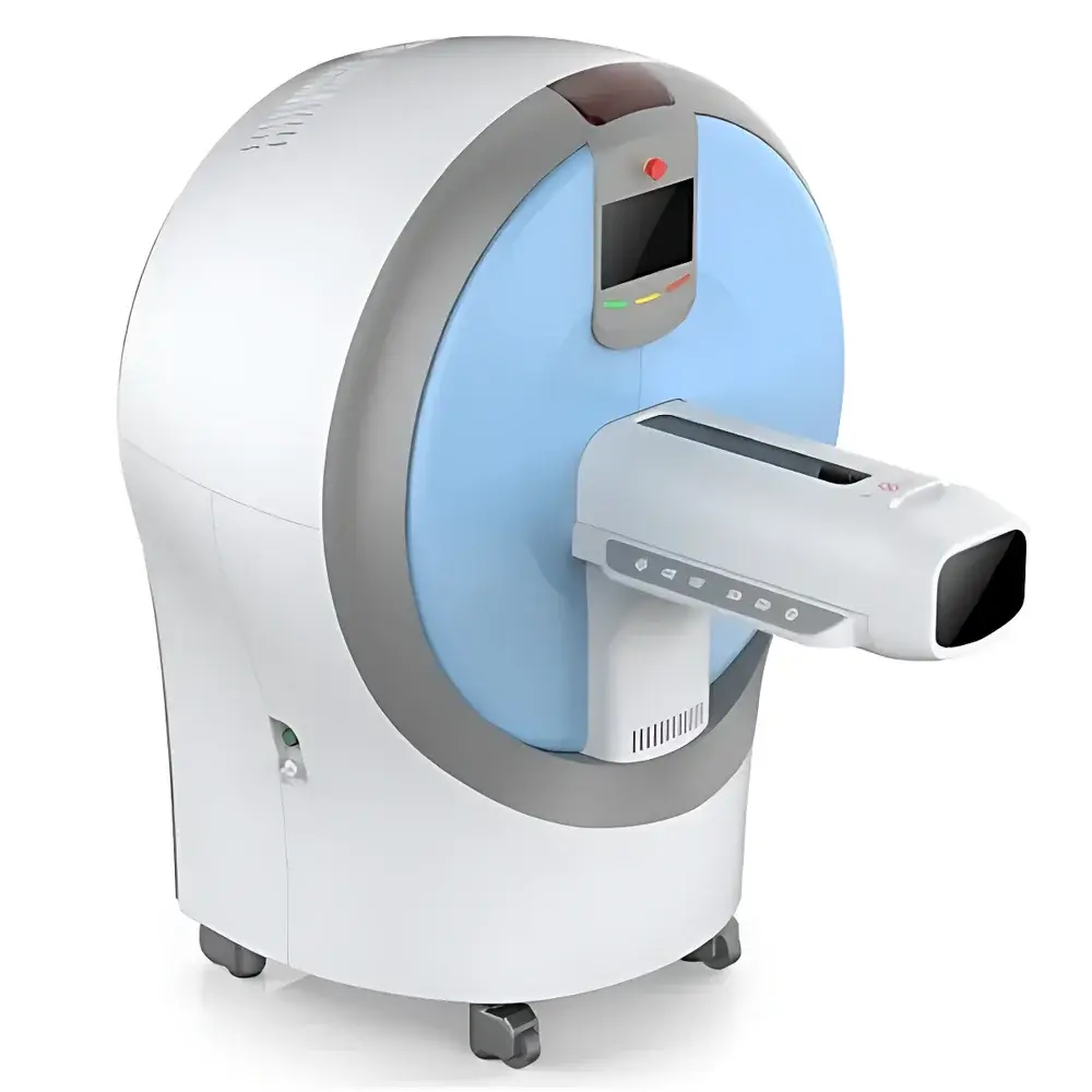
Aolong Small Animal Spectral Micro-CT System
| Brand | Aolong |
|---|---|
| Origin | Liaoning, China |
| Manufacturer Type | Original Equipment Manufacturer (OEM) |
| Country of Origin | China |
| Model | Small Animal Spectral Micro-CT |
| Field of View | Ø65 mm × L200 mm |
| X-ray Source | 90 kV sealed-tube, maintenance-free |
| Detector | Photon-counting energy-resolving detector |
| Minimum Pixel Resolution | 8 µm |
| Energy Bins | 2–8 selectable |
| Image Matrix | 512×512 to 1547×1547 |
| Radiation Leakage | <0.5 µSv/h at any external point |
| Dimensions (H×W×D) | 1622×1192×1458 mm |
| Power Supply | 100–240 VAC, 50–60 Hz, <300 W (excl. workstation) |
| Workstation | Intel Xeon 8-core CPU, 64 GB RAM, 256 GB SSD + 1 TB SSD + 4 TB HDD, 29″ display |
Overview
The Aolong Small Animal Spectral Micro-CT System is a preclinical computed tomography platform engineered for high-fidelity, quantitative 3D imaging of small animal models under in vivo and ex vivo conditions. Leveraging spectral (energy-resolved) X-ray detection technology, the system operates on the principle of multi-energy photon counting—capturing transmitted X-ray photons across discrete energy bins to enable material decomposition based on atomic number-dependent attenuation signatures. Unlike conventional broadband CT, this spectral architecture supports K-edge imaging, allowing selective enhancement of contrast agents containing elements such as iodine, gadolinium, or bismuth through targeted energy bin selection. The system delivers isotropic spatial resolution down to 8 µm with a cylindrical field of view of Ø65 mm × 200 mm in length—optimized for murine (mouse, rat), rabbit, and avian models—and integrates a 90 kV sealed microfocus X-ray source designed for long-term stability and minimal maintenance.
Key Features
- Energy-resolving photon-counting detector enabling quantitative material decomposition and virtual monoenergetic image reconstruction
- K-edge imaging capability for targeted contrast agent visualization and quantification (e.g., iodine, ytterbium, gold nanoparticles)
- High-contrast, high-resolution tomographic imaging optimized for soft-tissue differentiation—even among tissues with similar electron density (e.g., muscle vs. tumor stroma)
- Dedicated scanning protocols preconfigured for common preclinical models: trabecular bone analysis, lung parenchyma assessment, vascular perfusion studies, and longitudinal tumor growth monitoring
- Modular, interchangeable animal beds with integrated anesthesia gas ports compatible with standard rodent ventilators and vaporizers; rapid bed swap enables seamless transition between in vivo and ex vivo workflows
- Full radiation-shielded enclosure meeting IEC 61331-1 compliance, with measured ambient dose rate <0.5 µSv/h at all external surfaces
- Native support for multimodal image fusion via DICOM-RT and NIfTI interfaces—enabling co-registration with PET, SPECT, and optical imaging datasets
Sample Compatibility & Compliance
The system accommodates live or euthanized specimens ranging from neonatal mice to adult rats, guinea pigs, and chick embryos. Anesthesia-compatible positioning systems ensure physiological stability during acquisition, minimizing motion artifacts while maintaining compliance with institutional animal care and use committee (IACUC) guidelines. All hardware and software components are designed in accordance with ISO 13485 principles for medical device quality management and align with GLP-compliant data integrity requirements. Radiation safety performance conforms to IEC 62464-1 (preclinical imaging equipment) and national regulatory standards for Class II X-ray devices. Data acquisition logs—including tube voltage, current, exposure time, energy bin configuration, and gantry rotation parameters—are automatically embedded in DICOM headers to support auditability and traceability per FDA 21 CFR Part 11 expectations.
Software & Data Management
The proprietary acquisition and reconstruction suite provides real-time preview, iterative reconstruction (including ordered-subset expectation maximization), and GPU-accelerated 3D volume rendering. Material decomposition algorithms generate quantitative maps of constituent mass fractions (e.g., calcium hydroxyapatite, water, lipid, iodine) in standardized units (mg/cm³). Export formats include DICOM-CT, NIfTI, TIFF stacks, and STL for downstream analysis in MATLAB, Amira, Avizo, or commercial platforms such as Bruker’s CTAn or Skyscan’s CTVox. The bundled workstation features dual SSD storage tiers (OS + cache) and enterprise-grade HDD archiving (4 TB), supporting automated backup to network-attached storage (NAS) or PACS environments. Audit trails record user login, parameter changes, reconstruction settings, and export events—enabling full reproducibility and regulatory readiness.
Applications
- Longitudinal osteoporosis studies: quantification of trabecular thickness, spacing, and bone mineral density (BMD) with sub-10 µm morphometric precision
- Pulmonary phenotyping: airway lumen segmentation, emphysema index calculation, and alveolar surface area estimation in COPD and fibrosis models
- Oncology research: tumor volume tracking, necrotic core delineation, and contrast-enhanced vascular permeability mapping
- Cardiovascular development: 4D cardiac gating for murine heart functional analysis (ejection fraction, wall thickening)
- Biomaterial integration: scaffold degradation kinetics and host tissue ingrowth assessment in orthopedic and dental implant models
- Multimodal validation: structural correlation of CT-derived anatomy with functional PET/SPECT tracer uptake or bioluminescent signal distribution
FAQ
What is the minimum achievable voxel size in high-resolution mode?
The system achieves an isotropic voxel size of 8 µm under optimal magnification and geometric configuration, contingent upon sample size, source-to-detector distance, and reconstruction kernel selection.
Does the system support respiratory and cardiac gating?
Yes—integrated physiological monitoring inputs accept analog signals from pressure pads or ECG leads to trigger prospective or retrospective gating during acquisition.
Can spectral data be exported for custom material decomposition?
Yes—raw energy-bin projection data and calibrated detector response curves are accessible via API for third-party algorithm development and validation.
Is the workstation included in the base configuration?
Yes—the system ships with a validated, pre-configured workstation meeting specified hardware requirements, including RAID-protected storage and dual-monitor support.
How is radiation dose monitored and controlled during in vivo scans?
Dose is calculated in real time using Monte Carlo–based simulation models embedded in the acquisition software; users may set dose limits per scan or per timepoint, with automatic exposure modulation to maintain target SNR within constraint boundaries.
Related Products


