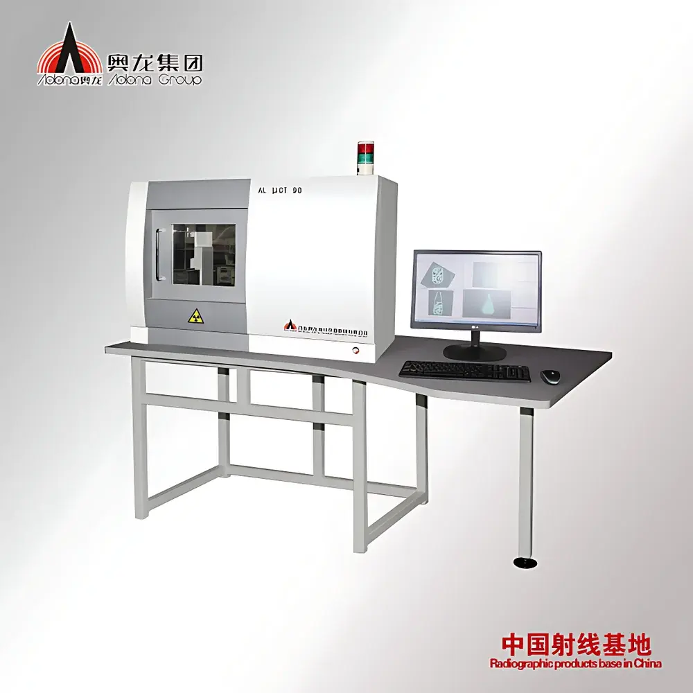
Aolong SmallFocus Micro-Focus X-ray Imaging System for Small Animals and Aquatic Specimens
| Brand | Aolong |
|---|---|
| Origin | Liaoning, China |
| Manufacturer Type | Direct Producer |
| Country of Origin | China |
| Model | SmallFocus |
| Pricing | Available Upon Request |
| Tube Voltage Range | 20–90 kV |
| Focal Spot Size | 5 µm |
| Active Imaging Area | 325 mm × 325 mm |
| Flat-Panel Detector Resolution | 100 µm |
| Chamber Material | Stainless Steel |
| Exposure Control | Fully Automated One-Touch Exposure |
| Remote Diagnostics | Integrated Ethernet-Based Support |
| Image Processing | Dedicated Workstation Included |
Overview
The Aolong SmallFocus Micro-Focus X-ray Imaging System is a dedicated benchtop radiographic platform engineered for high-resolution, non-invasive structural imaging of small living specimens—including laboratory rodents (mice, rats), lagomorphs (rabbits), companion animals (dogs, cats), non-human primates, ovine neonates, and aquatic models such as zebrafish and medaka. Unlike conventional diagnostic X-ray systems, the SmallFocus employs micro-focus X-ray tube technology with a nominal focal spot size of 5 µm, enabling geometric magnification imaging with sub-100 µm effective spatial resolution under optimized geometry. Its operation is grounded in transmission radiography: X-rays generated at adjustable tube voltages (20–90 kV) pass through biological tissue, where differential attenuation—governed by atomic number, density, and thickness—produces contrast in the digital radiograph. The system is designed for longitudinal in vivo studies requiring repeatable, low-dose imaging protocols compliant with ALARA (As Low As Reasonably Achievable) principles.
Key Features
- Micro-focus X-ray source (5 µm focal spot) delivering high spatial resolution and minimal penumbral blurring for magnified imaging
- Fully automated exposure control: one-touch acquisition eliminates dependency on operator expertise in radiographic technique selection
- Stainless steel imaging chamber with seamless welds and chemical-resistant surface—designed for rapid decontamination between subjects and compatibility with veterinary disinfectants
- Digital flat-panel detector with native pixel pitch of 100 µm and active area of 325 mm × 325 mm, supporting both contact and geometrically magnified imaging modes
- Integrated Ethernet interface enabling remote diagnostics, firmware updates, and real-time technical support without on-site service visits
- Dedicated image processing workstation pre-installed with quantitative analysis tools including ROI-based densitometry, edge-enhanced segmentation, and DICOM-compliant export
- No external lead shielding required: self-contained cabinet meets IEC 62464-1:2017 radiation safety limits for Class II cabinet X-ray systems when operated within specified voltage and exposure time parameters
Sample Compatibility & Compliance
The SmallFocus accommodates specimens ranging from 3-mm zebrafish larvae to 15-kg juvenile canines or capuchin monkeys. Adjustable subject positioning stages support lateral, dorsoventral, and oblique projections. For aquatic specimens, optional water-filled acrylic cradles maintain physiological hydration during imaging without compromising X-ray transmission. The system conforms to ISO 15801:2015 (non-destructive testing — industrial computed tomography) for image quality assurance and supports audit-ready documentation per GLP (Good Laboratory Practice) requirements. While not FDA-cleared for human clinical use, its design aligns with ASTM F2993-15 standards for preclinical imaging device performance validation.
Software & Data Management
Acquisition and post-processing are managed via Aolong’s proprietary SmallFocus Imaging Suite, a Windows-based application compliant with DICOM 3.0 Part 10 file structure. The software includes dose logging per acquisition, user-access-level controls (administrator, technician, trainee), and full audit trail functionality—including timestamps, operator ID, exposure parameters, and image modification history—to satisfy 21 CFR Part 11 electronic record integrity requirements. Raw projection data and reconstructed images are stored in encrypted local repositories with optional PACS integration via DICOM SCU/SCP protocols. Batch processing supports standardized measurement pipelines for bone mineral density (BMD) estimation, soft-tissue lesion volumetry, and developmental morphometric tracking across longitudinal cohorts.
Applications
- Longitudinal monitoring of orthopedic disease progression (e.g., osteoarthritis, fracture healing) in murine models
- Cardiovascular phenotyping: visualization of calcified plaques, cardiac chamber dimensions, and thoracic vasculature in genetically modified rats
- Developmental biology: skeletal ossification staging in zebrafish embryos and larval stages
- Oncology research: detection and volumetric quantification of subcutaneous and orthotopic tumors
- Toxicology screening: assessment of pulmonary edema, gastric distension, or renal calcification following compound administration
- Veterinary translational studies: pre-surgical planning and post-operative evaluation in companion animal patients
FAQ
Is the SmallFocus system suitable for regulatory submission studies?
Yes—when operated under documented SOPs and with full audit trail enabled, image datasets meet evidentiary requirements for ICH M3(R2), OECD 407, and FDA guidance on nonclinical imaging endpoints.
Can the imaging chamber accommodate live fish without anesthesia?
Yes—water-immersion imaging is supported using temperature-stabilized acrylic holders; motion artifacts are mitigated via ultra-short exposure times (< 100 ms) at higher kV settings.
What maintenance is required for the micro-focus X-ray tube?
The sealed metal-ceramic tube has a rated lifetime of ≥ 5,000 hours; routine maintenance is limited to annual calibration verification and detector flat-field correction.
Does the system support 3D reconstruction?
No—SmallFocus is a 2D projection radiography platform. For tomographic applications, Aolong offers the complementary MicroCT Focus series with cone-beam geometry and filtered back-projection reconstruction.
How is radiation safety ensured for daily lab use?
The stainless steel cabinet attenuates leakage radiation to < 0.5 µSv/h at 5 cm from any surface during operation, satisfying national regulations for unshielded cabinet X-ray devices in academic and pharmaceutical research facilities.


