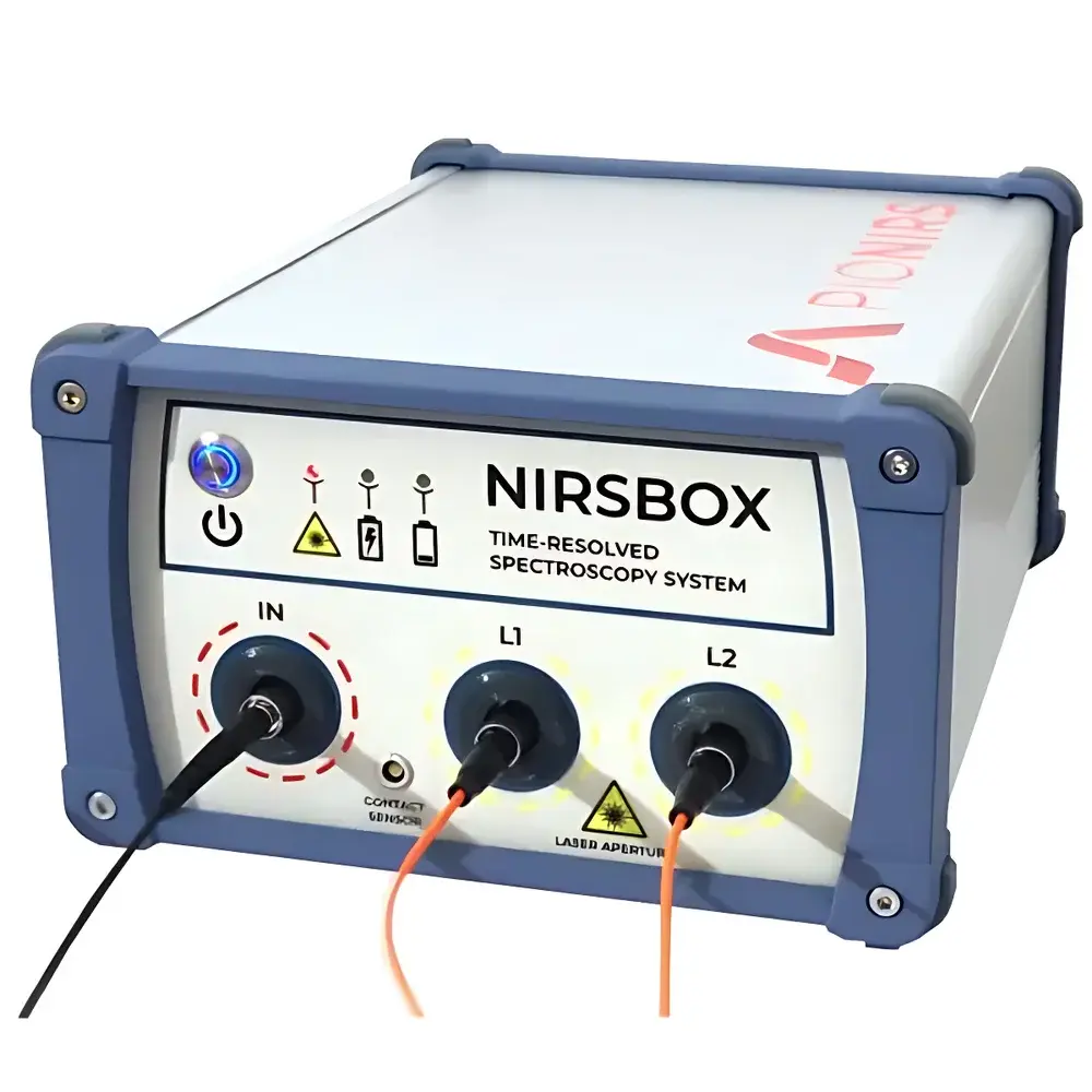
Auniontech NIRSBOX TD-NIRS Time-Domain Near-Infrared Spectrometer
| Brand | Auniontech |
|---|---|
| Origin | Shanghai, China |
| Manufacturer Type | Authorized Distributor |
| Product Category | Domestic |
| Model | NIRSBOX |
| Spectral Sensor Type | Time-Domain Photon Counting Detector |
| Spectral Range | 650–950 nm (Near-Infrared) |
| Interface | USB 2.0 |
| Output | Analog voltage proportional to physiological parameters |
| Compliance | MEDPHOT Protocol Verified |
| Stability | <1% drift over 14 h |
| Reproducibility | <2% monthly variation in μₐ and μₛ′ on tissue-mimicking phantoms |
Overview
The Auniontech NIRSBOX TD-NIRS Time-Domain Near-Infrared Spectrometer is a compact, dual-wavelength, time-resolved optical instrument engineered for quantitative functional monitoring of biological tissues. Based on time-domain near-infrared spectroscopy (TD-NIRS), the system measures the temporal distribution of photons—specifically the time-of-flight (ToF) histogram—after propagation through scattering media such as brain or muscle tissue. This principle enables absolute quantification of absorption (μₐ) and reduced scattering (μₛ′) coefficients without reliance on empirical calibration or spatial assumptions. Unlike continuous-wave (CW) or frequency-domain (FD) systems, TD-NIRS provides intrinsic depth sensitivity via photon arrival time gating, allowing depth-resolved hemodynamic parameter retrieval—including absolute concentrations of oxyhemoglobin ([HbO₂]), deoxyhemoglobin ([HbR]), total hemoglobin ([tHb]), and tissue oxygen saturation (StO₂)—with high reproducibility and minimal inter-subject variability.
Key Features
- Time-resolved photon detection with picosecond timing resolution using custom single-photon avalanche diode (SPAD) modules and time-correlated single-photon counting (TCSPC) electronics
- Dual-wavelength operation at 760 nm and 850 nm—optimized for differential hemoglobin spectroscopy and minimization of water interference
- Plug-and-play USB 2.0 interface enabling direct integration with Windows, Linux, or macOS platforms without external power supplies or warm-up delays
- Long-term measurement stability: <1% intensity drift over 14-hour continuous operation under ambient laboratory conditions
- Validated against the MEDPHOT protocol for diffuse optical instrumentation—ensuring traceability to international performance benchmarks for μₐ and μₛ′ recovery
- Analog output channel delivering real-time voltage signals linearly proportional to [HbO₂], [HbR], or StO₂—synchronized with external triggers via TTL I/O ports
Sample Compatibility & Compliance
The NIRSBOX is designed for non-invasive, contact-based interrogation of human and animal tissues including cerebral cortex, skeletal muscle, and superficial adipose layers. Its optical probe configuration supports standard source–detector separations (2–4 cm), compatible with both commercial optode holders and custom headgear. All hardware and firmware comply with IEC 61000-6-3 (EMC emission standards) and IEC 60601-1 (general safety requirements for medical electrical equipment). While classified as a research-use-only (RUO) device, its data acquisition architecture supports GLP/GMP-aligned workflows: raw ToF histograms are timestamped with microsecond precision; metadata (wavelength, gain, integration time) is embedded in HDF5-formatted output files for audit-ready traceability.
Software & Data Management
NIRSBOX Control Software (v3.x, Windows/Linux) provides real-time visualization of photon time-of-flight distributions, dynamic hemodynamic parameter maps, and export of calibrated physiological time series. The software implements standardized inverse modeling algorithms—including convolution-based fitting of the diffusion equation impulse response—to retrieve μₐ and μₛ′ from measured ToF curves. Raw data exports support MATLAB (.mat), Python-compatible HDF5, and CSV formats. For regulated environments, optional software validation packages include IQ/OQ documentation templates and 21 CFR Part 11-compliant user access logs, electronic signatures, and audit trail generation—facilitating integration into clinical trial or preclinical study data management systems.
Applications
- Functional brain imaging during cognitive tasks, neurofeedback training, or sleep studies—leveraging depth-resolved cortical activation mapping
- Monitoring muscle oxygenation kinetics during exercise physiology protocols or rehabilitation interventions
- Neonatal cerebral oximetry with motion-robust artifact rejection and low-SNR tolerance
- Preclinical longitudinal studies in rodent models of stroke, traumatic brain injury, or tumor metabolism
- Method development for multimodal integration—e.g., simultaneous acquisition with EEG, fMRI, or diffuse correlation spectroscopy (DCS)
- Calibration reference for CW-NIRS systems requiring absolute baseline correction
FAQ
What optical properties can be quantified directly from TD-NIRS measurements?
Absorption coefficient (μₐ) and reduced scattering coefficient (μₛ′) are retrieved by fitting the measured photon time-of-flight distribution to solutions of the time-dependent radiative transfer equation—enabling absolute, wavelength-specific quantification without reference measurements.
Is the NIRSBOX suitable for clinical use?
The NIRSBOX is designated for research use only (RUO); it is not CE-marked or FDA-cleared for diagnostic or therapeutic applications. However, its MEDPHOT-validated performance supports translational study design and regulatory submission preparation.
How is synchronization achieved with external devices like EEG or DCS systems?
The instrument provides TTL-compatible trigger input and output ports, supporting hardware-level synchronization with sub-millisecond jitter—essential for multimodal event-locked analysis.
Can raw time-of-flight histograms be exported for custom processing?
Yes. Full-resolution ToF histograms (up to 1024 time bins per wavelength) are saved in HDF5 format with embedded metadata, enabling offline reconstruction using open-source toolkits such as NIRX or Homer3.
What is the typical source–detector separation supported?
The standard fiber-optic probe configuration supports 30 mm and 40 mm separations; custom probes with 20–50 mm spacing are available upon request for depth-specific interrogation.