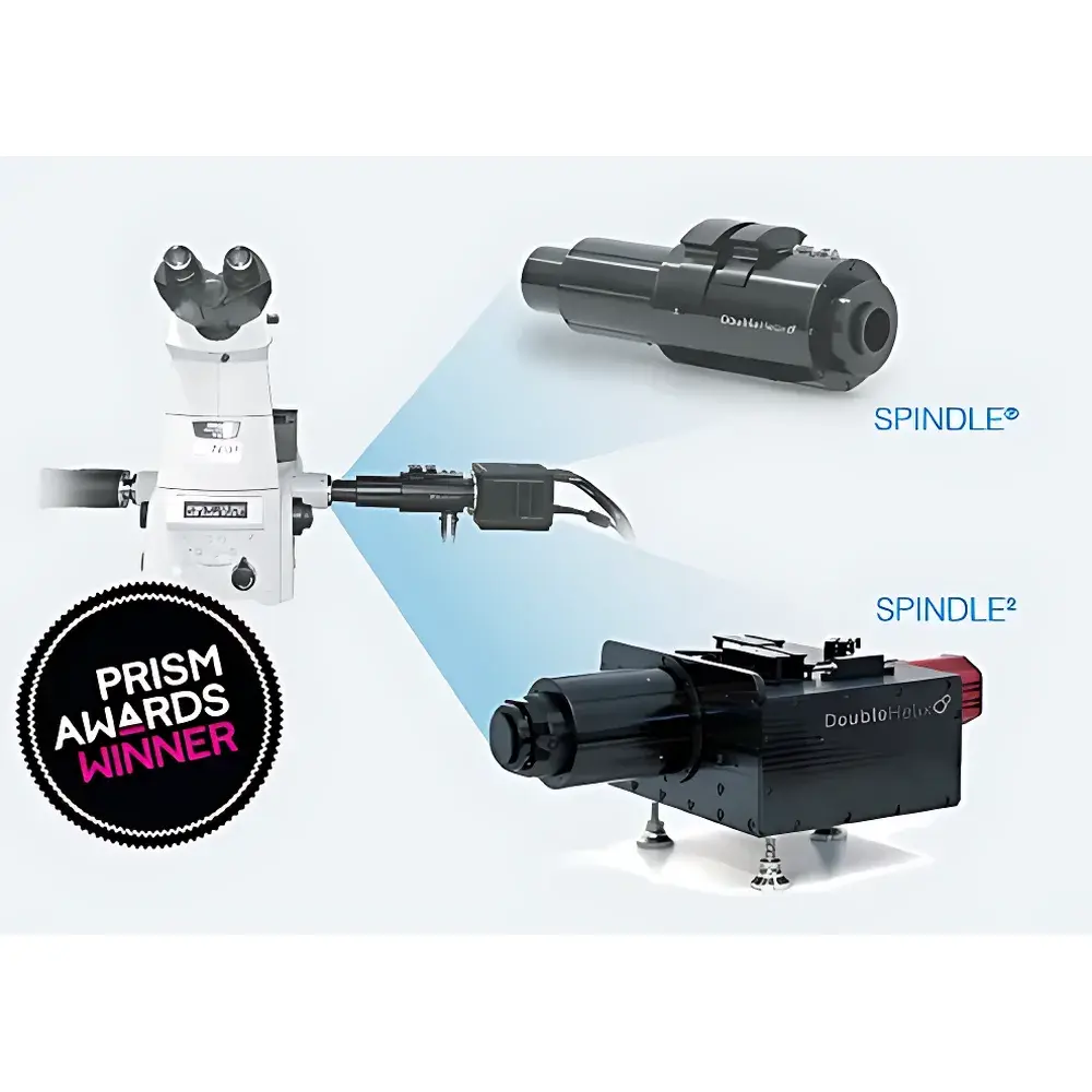
Auniontech SPINDLE 3D Super-Resolution Single-Molecule Localization Microscopy Module (Scan-Free)
| Brand | Auniontech |
|---|---|
| Model | SPINDLE |
| Origin | Imported |
| Dimensions | 210 mm × 84 mm × 84 mm |
| Depth Range | 2.2 µm |
| Transmission Efficiency | 95% |
| Wavelength Range | 400 nm – NIR |
| Compatible Detectors | EMCCD & sCMOS cameras |
| Interface | Standard C-mount |
| Software | 3DTRAX for 3D localization, reconstruction, and tracking |
Overview
The Auniontech SPINDLE 3D Super-Resolution Single-Molecule Localization Microscopy Module is an engineered optical add-on designed to convert conventional widefield, TIRF, or light-sheet microscopes into high-precision, scan-free 3D super-resolution imaging platforms. Unlike scanning-based modalities such as confocal or STED, SPINDLE leverages engineered point spread function (E-PSF) optics—specifically a double-helix phase mask—to encode axial (z) position information directly into the lateral (x–y) intensity distribution of single-molecule emissions. This enables simultaneous acquisition of x, y, and z coordinates from a single camera frame, eliminating mechanical scanning and preserving temporal fidelity essential for live-cell dynamics. The module operates across a broad spectral range (400 nm to NIR), maintains >95% optical transmission, and delivers sub-diffraction resolution with typical lateral localization precision of ≤20 nm and axial precision of ≤25 nm over a calibrated depth range of up to 20 µm—achievable without hardware modification to the host microscope’s optical train.
Key Features
- Scan-free 3D single-molecule localization: Captures full 3D coordinates in a single exposure using double-helix E-PSF encoding.
- Modular integration: Installs between objective and camera via standard C-mount interface; compatible with EMCCD and sCMOS detectors.
- Interchangeable phase mask library: Enables optimization of depth range, axial sensitivity, and signal-to-noise ratio for specific fluorophores and experimental configurations.
- Bypass mode: Allows rapid reversion to native 2D widefield or TIRF imaging without physical disassembly.
- Compact form factor (210 × 84 × 84 mm): Designed for minimal footprint and vibration-insensitive alignment in multi-instrument setups.
- Calibrated depth range: Up to 20 µm with uniform localization precision; configurable depth tuning from 2.2 µm upward based on mask selection.
Sample Compatibility & Compliance
SPINDLE supports fixed and live biological specimens—including whole cells, membrane proteins, cytoskeletal structures, and nucleic acid complexes—under standard fluorescence labeling protocols (e.g., Alexa Fluor, ATTO, Cy dyes). It is fully compatible with established super-resolution modalities including STORM, PALM, SOFI, and FRET, and integrates seamlessly with light-sheet and multi-color widefield systems. For regulated environments, the 3DTRAX software architecture supports audit trails, user access controls, and metadata logging aligned with GLP/GMP documentation requirements. While SPINDLE itself is not a medical device, its use in research workflows adheres to ISO/IEC 17025 principles for measurement traceability, and data export formats (TIFF, HDF5, CSV) conform to FAIR data standards for long-term archival and cross-platform analysis.
Software & Data Management
The proprietary 3DTRAX software suite provides end-to-end processing for 3D single-molecule localization microscopy: real-time drift correction, PSF fitting (Gaussian or double-helix model), 3D reconstruction, trajectory linking, and quantitative analysis (e.g., cluster density, diffusion coefficient, co-localization statistics). All processing steps are scriptable via Python API, enabling reproducible pipeline execution and integration into automated analysis workflows. Raw image data retain full bit-depth fidelity; localization tables include uncertainty estimates for each coordinate (σx, σy, σz) and photon count metrics. Optional modules include deconvolution-enhanced volumetric rendering and extended-depth-of-field synthesis for whole-cell context mapping—particularly valuable when correlating nanoscale molecular distributions with cellular ultrastructure.
Applications
- Multi-particle 3D tracking in live cells: Quantifying intracellular transport, vesicle trafficking, and chromatin dynamics with millisecond temporal resolution.
- Multi-color SMLM: Simultaneous 3D mapping of ≥4 spectrally distinct targets using SPINDLE2 dual-channel configuration.
- Whole-cell + single-molecule correlative imaging: Combining diffraction-limited structural context with nanoscale molecular annotation.
- Light-sheet compatibility: Enabling volumetric super-resolution in cleared tissues or organoids without axial scanning artifacts.
- Single-molecule counting and stoichiometry analysis: Leveraging precise z-localization to resolve overlapping emitters in dense clusters.
- Structural biology applications: Resolving conformational heterogeneity in membrane protein assemblies and nucleoprotein complexes.
FAQ
Does SPINDLE require modification to my existing microscope?
No—SPINDLE installs as a drop-in module between the microscope output port and camera, requiring no optical realignment or permanent alterations.
Can SPINDLE be used with TIRF or light-sheet systems?
Yes—its passive, alignment-tolerant design supports integration with TIRF, HILO, and orthogonal light-sheet geometries without compromising evanescent field integrity or illumination uniformity.
What camera specifications are recommended for optimal performance?
EMCCD cameras with quantum efficiency >90% at target emission wavelengths and sCMOS cameras with ≤1.5 e⁻ read noise and ≥82% QE are strongly recommended; pixel size should be matched to system magnification for Nyquist sampling of the engineered PSF.
Is 3DTRAX validated for regulatory submissions?
While 3DTRAX is not FDA-cleared, it includes features supporting 21 CFR Part 11 compliance (electronic signatures, audit logs, version-controlled processing scripts) for preclinical research documentation.
How is calibration performed for axial localization accuracy?
Calibration uses fluorescent nanospheres immobilized in polyacrylamide gels at known depths; the 3DTRAX calibration workflow generates per-wavelength, per-magnification PSF models with quantified z-dependent bias and precision metrics.


