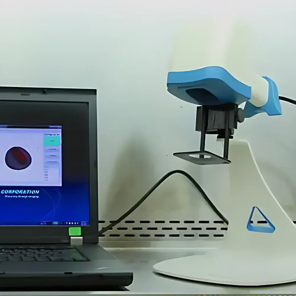
Biopticon TumorImager 2 Small Animal Subcutaneous Tumor Imaging and Measurement System
| Brand | Biopticon |
|---|---|
| Origin | USA |
| Manufacturer Type | Authorized Distributor |
| Product Category | Imported Instrument |
| Model | TumorImager 2 |
| Instrument Type | Optical 3D Surface Imaging System |
| Scan Resolution | 480 × 640 pixels |
| Scan Speed | <1 s per acquisition |
| Field of View | 30 mm × 20 mm |
| Sample Capacity | 1 animal per scan |
| Z-Axis Resolution | 50 µm |
| Interface | USB 3.0 |
| Output Formats | JPG (2D), OBJ & PLY (3D surface mesh) |
| Operating Modes | Handheld or Stand-Mounted |
| Tumor Length Limit | ≤30 mm |
| Tumor Height Limit | ≤20 mm |
| Automatic Segmentation Time | ~3 s |
| Data Storage | Local SQL database with user authentication and audit trail |
Overview
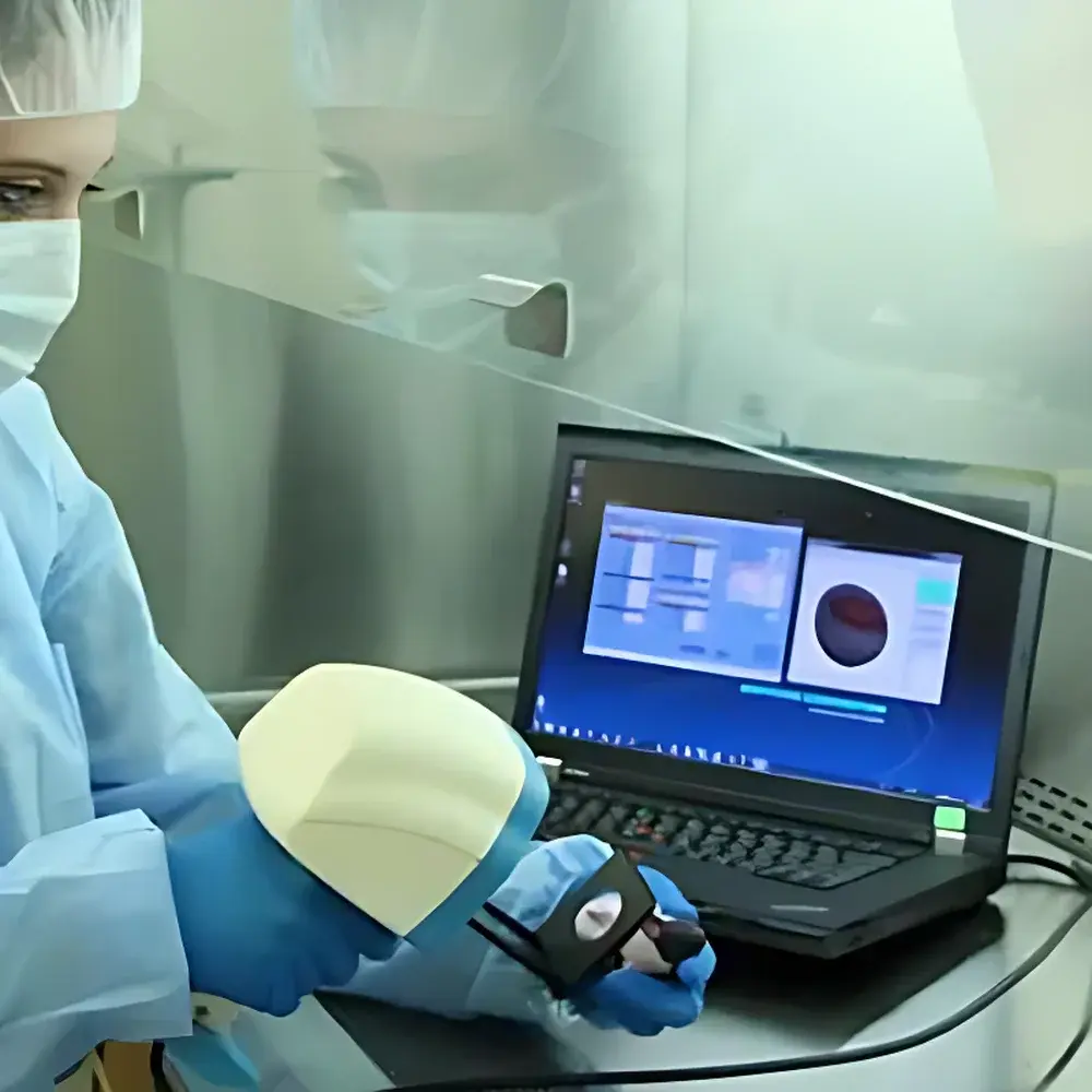
The Biopticon TumorImager 2 is a second-generation, non-invasive optical 3D surface imaging system engineered for precise, repeatable volumetric measurement of subcutaneous tumors in live rodents. Unlike conventional caliper-based estimation—subject to inter-operator variability, anatomical assumption bias (e.g., ellipsoid volume approximation), and limited sensitivity to subtle morphological changes—the TumorImager 2 employs structured-light laser scanning to reconstruct high-fidelity topographic surface profiles. This contactless, radiation-free method captures the true geometric contour of tumor masses in vivo without requiring anesthesia, sedation, or physical restraint beyond gentle manual positioning. The system operates on the principle of triangulation-based profilometry: a calibrated laser line projects across the tumor surface while a synchronized CMOS sensor records displacement-induced distortions in the line profile, enabling sub-50 µm axial resolution reconstruction. Designed specifically for preclinical oncology workflows, it delivers objective, operator-independent quantitative metrics—including absolute volume (mm³), surface area (mm²), longitudinal growth kinetics, tumor doubling time (TDT), and log cell kill estimates—directly traceable to GLP-compliant experimental records.
Key Features
- Contactless, anesthesia-free acquisition: Eliminates physiological stress artifacts and reduces procedural burden on animal subjects and technicians.
- Dual operational modes: Switch seamlessly between handheld scanning for flexibility during longitudinal studies and rigid stand-mounted configuration for standardized positional repeatability.
- Sub-second acquisition (<1 s) with real-time 3D visualization: Enables rapid throughput in multi-animal cohorts without compromising data fidelity.
- Adaptive tumor segmentation: Supports both automated threshold-based contour detection and manual refinement via touchscreen, mouse, or footswitch—critical for heterogeneous, necrotic, or irregularly shaped lesions.
- True 3D volumetric quantification: Generates exportable OBJ and PLY mesh files for downstream morphometric analysis, co-registration with MRI/CT datasets, or archival in institutional digital repositories.
- Integrated hardware-software synchronization: USB 3.0 interface ensures deterministic latency control and lossless transfer of raw scan data to the TumorManager platform.
Sample Compatibility & Compliance
The TumorImager 2 is validated for use with murine models (C57BL/6, BALB/c, nude, NSG) bearing subcutaneous xenografts, syngeneic, or genetically engineered tumors. It accommodates tumors up to 30 mm in length and 20 mm in height—covering >95% of standard preclinical tumor burden ranges. No contrast agents, radioactive tracers, or surgical preparation are required. The system complies with ISO 13485–aligned quality management protocols for research instrumentation and supports 21 CFR Part 11–compliant electronic records through role-based access control, password-protected database encryption, and full audit trail logging of all user actions (scan initiation, segmentation edits, report generation). All measurement algorithms adhere to ASTM E2917-22 guidelines for uncertainty estimation in biomedical dimensional metrology.
Software & Data Management
TumorManager is a dedicated, SQL-backed desktop application that transforms raw scans into auditable experimental outputs. It enforces structured metadata capture—including animal ID, strain, treatment group, dosing regimen, and observer credentials—at point-of-acquisition. Volume calculations apply validated geometric integration over reconstructed surface meshes—not empirical formulas—ensuring traceability and reproducibility across laboratories. Statistical modules compute intra-group variance, inter-group significance (unpaired t-test, ANOVA), and longitudinal growth curve fitting (exponential, Gompertz). Reports export natively to PDF, Excel, and Word formats with embedded charts, metadata headers, and customizable templates compliant with ICH-GCP and NIH grant reporting standards. Database backups are automated and version-stamped; historical scans remain searchable by date, operator, or phenotype tag.
Applications
- Preclinical efficacy evaluation of cytostatic and cytotoxic oncology therapeutics (monotherapies and combinations).
- Pharmacodynamic biomarker correlation studies linking tumor volume kinetics to molecular endpoints (e.g., Ki67, cleaved caspase-3).
- Validation of orthotopic or metastatic models where subcutaneous surrogates serve as pharmacokinetic/pharmacodynamic bridges.
- Quality assurance in contract research organizations (CROs) performing GLP-compliant toxicology and safety pharmacology studies.
- Training platforms for standardizing tumor measurement practices across multi-site academic consortia.
FAQ
Does the TumorImager 2 require animal anesthesia during scanning?
No. The system is designed for conscious, gently restrained animals, eliminating confounding variables introduced by anesthetic agents on tumor perfusion and immune activity.
Can it accurately measure necrotic or ulcerated tumors?
Yes. Manual segmentation mode allows precise delineation of viable margins, and surface mesh reconstruction accommodates irregular topographies including central necrosis and epidermal disruption.
How does its volumetric accuracy compare to caliper measurements?
Independent validation studies show mean absolute error of ±0.1 mm³ versus ±0.36 mm³ for digital calipers under identical conditions—representing a >60% reduction in systematic bias and improved statistical power in dose-response analyses.
Is TumorManager compatible with LIMS or institutional ELN systems?
Yes. The software supports ODBC-compliant database exports and provides RESTful API endpoints for bidirectional integration with enterprise research informatics platforms.
What regulatory documentation is provided for audit readiness?
Each system ships with a Factory Acceptance Test (FAT) report, IQ/OQ documentation templates, and a Certificate of Conformance to ISO/IEC 17025 calibration traceability standards for the laser profiling subsystem.


