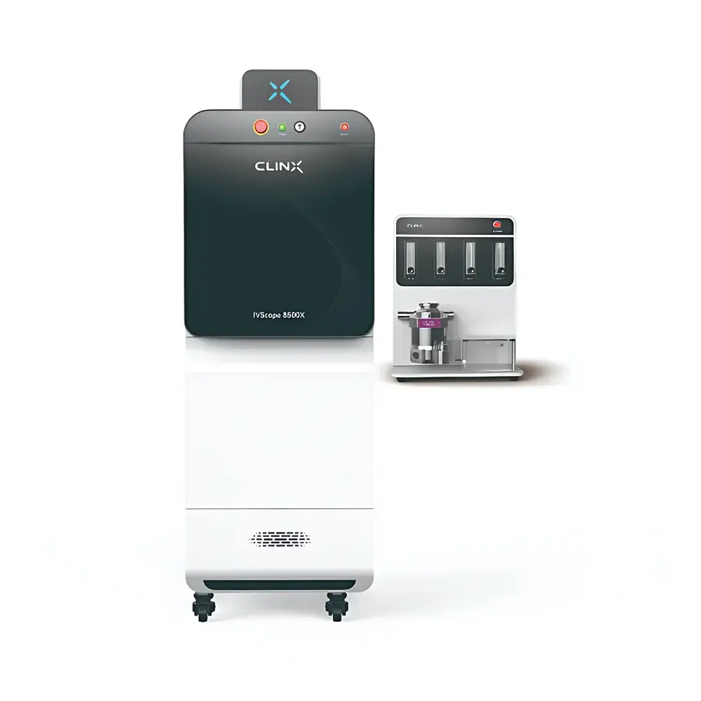
Clinx IVScope 8200X Multi-Modal X-ray and Optical Small Animal In Vivo Imaging System
| Brand | Clinx |
|---|---|
| Origin | Shanghai, China |
| Model | IVScope 8200X |
| Imaging Modality | Optical (Bioluminescence/Fluorescence) + X-ray |
| Max Sample Capacity | 5 mice |
| CCD Resolution | 1024 × 1024 |
| Field of View | 11 cm × 11 cm to 25 cm × 25 cm |
| X-ray Minimum Resolvable Feature Size | 0.16 mm |
| Lens Aperture | f/0.8 motorized auto-focus lens |
| Camera | 6 MP front-illuminated cooled CCD (optional water-cooling to −90 °C) |
| Sample Stage Temperature Control | Ambient to 42 °C |
| Radiation Shielding | ≤0.5 mR/h at 5 cm from base enclosure (compliant with FDA 21 CFR 1020.40) |
| Illumination Sources | Standard white-light reflectance |
| Filter System | Motorized filter wheel with emission filter slots |
| Anesthesia Interface | Integrated gas anesthesia delivery system |
| Software | FDA 21 CFR Part 11–compliant acquisition and quantification suite |
Overview
The Clinx IVScope 8200X is a preclinical multi-modal in vivo imaging platform engineered for simultaneous high-fidelity X-ray radiography and optical bioluminescence/fluorescence imaging in small animal models. It operates on dual physical principles: X-ray absorption contrast for anatomical localization and structural assessment, and photon detection via a thermoelectrically cooled, low-noise CCD sensor for functional molecular imaging. The system enables longitudinal, non-invasive monitoring of disease progression, therapeutic efficacy, and pharmacokinetic biodistribution—critical for oncology, immunology, neurodegenerative, and cardiovascular research. Its integrated design ensures spatial co-registration between X-ray and optical datasets without mechanical repositioning, eliminating registration error and enabling pixel-accurate overlay analysis. All radiation-emitting components comply with U.S. FDA 21 CFR 1020.40 for diagnostic X-ray equipment safety, including strict limits on leakage radiation and interlocked exposure control.
Key Features
- Multi-modal acquisition architecture: Synchronized acquisition of X-ray radiographs and bioluminescent/fluorescent signals within a single imaging session, supporting spectral unmixing and anatomical-functional fusion.
- Ultra-low-noise optical detection: 6-megapixel front-illuminated CCD sensor with thermoelectric cooling down to −90 °C (with optional water-circulation chiller), achieving dark current <0.001 e⁻/pixel/sec and enabling detection of sub-picomolar luciferase activity.
- f/0.8 motorized lens assembly: Large-aperture, autofocus optical train optimized for photon-limited conditions—maximizing signal capture across visible and near-infrared wavelengths (400–900 nm).
- Adjustable field-of-view (FOV): Continuously variable FOV from 11 cm × 11 cm (high-resolution mode) to 25 cm × 25 cm (whole-body mouse imaging), achieved via precision motorized zoom and stage positioning.
- Regulatory-compliant radiation management: Lead-shielded chamber with interlocked door sensors, real-time dose monitoring, and SAR shielding verified at ≤0.5 mR/h at 5 cm from enclosure walls per FDA 21 CFR 1020.40.
- Physiological support infrastructure: Integrated isoflurane vaporizer with flow metering, temperature-controlled stage (ambient to 42 °C), and motion-stabilized animal restraint trays for reproducible positioning across serial timepoints.
Sample Compatibility & Compliance
The IVScope 8200X accommodates live murine models (mouse, rat) weighing up to 50 g, with capacity for 1–5 animals per imaging session. Subjects are secured using modular, ventilated restraint platforms compatible with standard inhalation anesthesia tubing. All optical and X-ray protocols adhere to institutional animal care and use committee (IACUC) guidelines and the ARRIVE 2.0 reporting standards. The system supports GLP-compliant workflows through audit-trail-enabled software, electronic signatures, and secure user-role permissions aligned with FDA 21 CFR Part 11 requirements. X-ray exposure parameters (kVp, mAs, filtration) are preset according to ISO 15708-2 for small-animal radiography, and optical acquisition settings conform to ASTM E2597–21 for quantitative bioluminescence imaging.
Software & Data Management
Clinx ImageStudio Pro is the native acquisition and analysis suite, built on a modular, client-server architecture with DICOM 3.0 export capability. It provides real-time image preview, automatic background subtraction, spectral deconvolution for multiplexed fluorescent probes, and region-of-interest (ROI)-based quantification with coefficient-of-variation (CV) reporting. Raw data are stored in vendor-neutral HDF5 format with embedded metadata (exposure time, gain, binning, filter ID, anesthesia status, stage temperature). The software includes built-in tools for co-registration validation, dose mapping overlays, and batch processing pipelines compatible with MATLAB and Python (via documented REST API). Audit logs record all user actions—including parameter changes, ROI edits, and export events—with timestamped, immutable entries compliant with GLP/GMP documentation standards.
Applications
- Oncology: Longitudinal tracking of orthotopic tumor growth, metastasis, and response to checkpoint inhibitors or CAR-T therapy using dual BLI/X-ray fusion.
- Inflammation & Autoimmunity: Quantification of NF-κB reporter activity in arthritic joints correlated with soft-tissue swelling on X-ray.
- Cardiovascular Research: Assessment of myocardial infarction size via luciferase-labeled stem cell engraftment overlaid on cardiac silhouette from high-contrast radiography.
- Neuroscience: Monitoring of AAV-mediated gene expression in transgenic models using Cre-dependent luciferase reporters, spatially anchored to skull landmarks on X-ray.
- Drug Development: Biodistribution studies of NIR-II fluorophores conjugated to monoclonal antibodies, validated against anatomical reference from same-session X-ray.
FAQ
What X-ray safety certifications does the IVScope 8200X meet?
The system complies with FDA 21 CFR 1020.40 for cabinet X-ray systems, including maximum permissible leakage radiation (≤0.5 mR/h at 5 cm), interlocked exposure control, and warning indicators. It also meets IEC 62464-1 for medical X-ray equipment electromagnetic compatibility.
Can the system perform spectral unmixing for multiple fluorescent probes?
Yes—ImageStudio Pro supports linear unmixing of up to four spectrally distinct fluorophores using user-defined reference spectra and automated autofluorescence subtraction.
Is remote operation supported for core facility environments?
The system supports secure remote access via TLS-encrypted VNC and scheduled acquisition through the central server interface, with role-based authentication and session logging.
How is calibration maintained across imaging sessions?
Daily automated flat-field correction, weekly intensity reference checks using NIST-traceable LED sources, and quarterly X-ray beam homogeneity verification per ISO 15708-2 ensure longitudinal quantitative consistency.
Does the software support integration with LIMS or ELN systems?
Yes—HDF5 metadata export, DICOM-SR structured reporting, and REST API enable bidirectional integration with major laboratory informatics platforms including LabVantage, Benchling, and Dotmatics.
Related Products