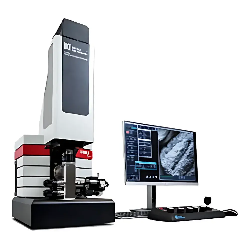
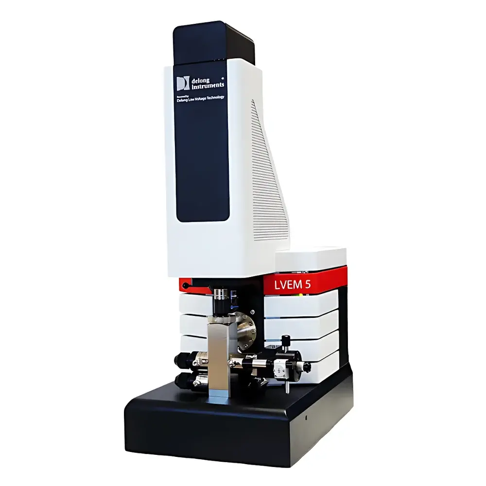
Delong LVEM5 Benchtop Low-Voltage Transmission Electron Microscope for Biological Applications
| Brand | Delong Instruments |
|---|---|
| Origin | Canada |
| Model | LVEM5 (Biological) |
| Accelerating Voltage | 5 kV |
| Maximum Magnification | 700,000× |
| Resolution (unstained biological samples) | 1.5 nm |
| Imaging Modes | TEM, SEM, STEM |
| Vacuum Pump-Down Time | ≤3 min |
| Footprint | <1 m² |
| Operating Environment | Standard laboratory (no external cooling, no high-vacuum infrastructure required) |
Overview
The Delong LVEM5 is a compact, benchtop low-voltage transmission electron microscope engineered specifically for high-fidelity structural analysis of unstained biological specimens. Unlike conventional high-voltage TEMs operating at 80–300 kV, the LVEM5 employs a 5 kV electron beam, which significantly enhances elastic scattering cross-sections for light elements (C, N, O, H)—the primary constituents of proteins, nucleic acids, lipids, and extracellular vesicles. This physical principle enables intrinsic contrast generation without heavy-metal staining (e.g., uranyl acetate or phosphotungstic acid), preserving native conformation and minimizing beam-induced damage. Its integrated column design eliminates the need for liquid nitrogen cooling, oil diffusion pumps, or dedicated shielded rooms—allowing deployment in standard biosafety level 2 (BSL-2) or BSL-3 laboratories with standard 110/220 V AC power and ambient air conditioning. The system achieves a verified point resolution of 1.5 nm under unstained conditions, validated across peer-reviewed studies on exosomes, bacteriophages, liposomes, and enveloped viruses.
Key Features
- Low-voltage electron optics (5 kV): Maximizes mass-thickness contrast for organic materials while reducing radiolysis and charging artifacts.
- Triple-mode imaging capability: Seamless switching between transmission electron microscopy (TEM), scanning electron microscopy (SEM), and scanning transmission electron microscopy (STEM) within a single platform—enabling correlative topographic and internal structural analysis.
- Rapid vacuum readiness: Integrated turbomolecular pumping achieves operational vacuum (<1×10⁻⁵ Pa) in ≤3 minutes—eliminating overnight pump-down delays typical of conventional TEMs.
- Benchtop footprint & infrastructure independence: Occupies less than 1 m²; requires no external water cooling, high-current circuits, or RF-shielded rooms—compatible with P3 containment labs where space and HVAC constraints prohibit traditional TEM installation.
- Automated alignment & intelligent UI: Guided workflows for gun alignment, stigmator correction, and focus calibration reduce operator dependency; all functions accessible via touchscreen interface with context-sensitive help.
- Low-dose imaging protocols: Software-controlled beam blanking, dwell time modulation, and frame averaging support quantitative structural preservation for radiation-sensitive specimens.
Sample Compatibility & Compliance
The LVEM5 is optimized for direct imaging of hydrated or air-dried biological nanomaterials without fixation, dehydration, embedding, or staining. Validated specimen types include: exosomes (30–150 nm), liposomes (50–200 nm), bacteriophages (e.g., T4, λ), influenza virions, SARS-CoV-2 pseudovirions, DNA-protein complexes, and amyloid fibrils. Its operation conforms to GLP-aligned documentation standards, supporting audit-ready image metadata (timestamp, magnification, kV, detector gain, exposure time). While not certified for GMP manufacturing environments per FDA 21 CFR Part 11, its software supports user access control, electronic signatures, and immutable audit trails—facilitating compliance with ISO/IEC 17025 and ASTM E2819-22 (Standard Guide for TEM Sample Preparation of Biological Nanomaterials).
Software & Data Management
Acquisition and analysis are performed using Delong’s proprietary LVEM Control Suite v4.x, built on a deterministic real-time Linux kernel. The software provides: (1) synchronized multi-modal acquisition (TEM+STEM dual-beam capture), (2) non-linear denoising via deep learning-based reconstruction (optional module), (3) batch processing with customizable export templates (TIFF, MRC, DM4), and (4) metadata embedding compliant with FAIR principles (Findable, Accessible, Interoperable, Reusable). Raw data files include full instrument parameter logs, enabling retrospective reprocessing and method validation. Export formats integrate natively with open-source platforms including IMOD, UCSF ChimeraX, and cryoSPARC Lite for subtomogram averaging or 2D class averaging.
Applications
- Extracellular vesicle characterization: Distinguishing exosome morphology, membrane integrity, and cargo density without negative stain-induced aggregation or flattening.
- Virology & vaccine development: Visualizing capsid symmetry, envelope glycoprotein distribution, and budding intermediates in near-native states—critical for epitope mapping and neutralization assay support.
- Nanomedicine quality control: Rapid assessment of liposomal drug carrier size distribution, lamellarity, and encapsulation homogeneity.
- Structural biology screening: Pre-cryo-EM grid assessment of particle dispersion, ice thickness estimation, and preferred orientation detection.
- Teaching & core facility democratization: Enabling undergraduate students and non-TEM specialists to acquire publication-grade micrographs with minimal training.
FAQ
Does the LVEM5 require liquid nitrogen or external cooling systems?
No. The LVEM5 uses thermoelectrically stabilized electron optics and does not incorporate field emission guns or cryo-stages requiring cryogens.
Can it image frozen-hydrated samples?
Not natively. The LVEM5 operates under high-vacuum conditions incompatible with ice sublimation control; however, it accepts air-dried or critical-point-dried replicas of vitrified grids for rapid feasibility screening.
Is training provided for new users?
Yes. Delong offers on-site installation, 2-day operator certification workshops, and remote troubleshooting support with SLA-backed response times.
What sample preparation methods are recommended?
Carbon-coated 200-mesh copper grids; optional glow discharge for hydrophilicity enhancement. No staining, fixation, or sectioning required for most soft biological targets.
How is data integrity ensured for regulatory submissions?
All acquisitions generate timestamped, parameter-locked digital records with SHA-256 checksums; optional integration with LIMS via RESTful API enables traceability from image to experimental protocol.


