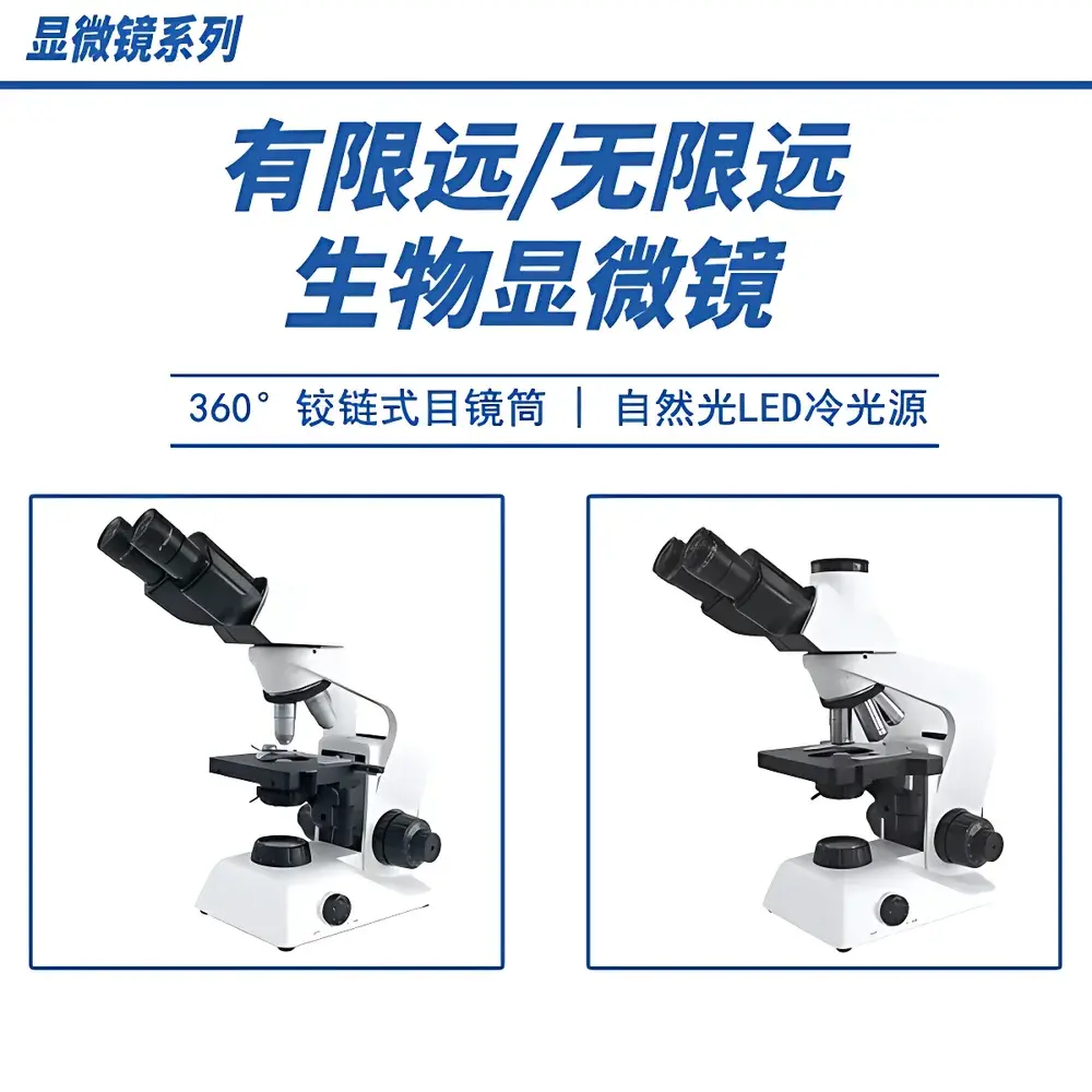
DS BH203 Biological Microscope
| Brand | DS |
|---|---|
| Origin | Beijing, China |
| Model | BH203 |
| Microscope Type | Upright Biological Microscope |
| Head Configuration | Trinocular |
| Optical System | LIOS Infinity-Corrected Achromatic Optical System |
| Magnification Range | 40×–1600× |
| Eyepieces | Wide-field, high-eyepoint plan eyepieces (10×, FN 20 mm |
| Objectives | Infinity-corrected achromatic objectives — 4×, 10×, 40× (spring-loaded), 100× oil (spring-loaded) |
| Interpupillary Adjustment | 48–75 mm (FN 20) / 48–76 mm (FN 22) |
| Nosepiece | Internal-facing, center-locating quadruple nosepiece (quintuple optional) |
| Focus Mechanism | Coaxial coarse/fine focus — coarse travel 26 mm, fine focus graduation 2 µm per division, 0.2 mm per revolution, upper limit stop and tension adjustment |
| Stage | Mechanical stage (142 × 133 mm), precision triangular guide rails, X/Y travel 76 × 52 mm, vernier scale 0.1 mm, dual-specimen clip, steel-wire X-axis drive, adjustable upper stop to prevent objective–specimen collision |
| Condenser | Abbe condenser, NA 1.25, iris diaphragm, vertical travel 12 mm |
| Illumination | 3 W LED cold light source (100–240 V AC input), continuously variable brightness, non-heat-emitting, high CRI, >25,000 h lifetime |
| Optional Accessories | Darkfield stop, polarizing kit (polarizer + analyzer), trinocular port with C-mount adapter for CMOS/CCD cameras (1×, 0.5×, 0.35× reduction options) |
Overview
The DS BH203 Biological Microscope is an upright, trinocular microscope engineered for undergraduate and graduate life science education, routine histology, and basic research applications requiring reliable optical performance and ergonomic operability. It employs the LIOS infinity-corrected achromatic optical system—a design standard in modern educational and entry-level research microscopes—ensuring consistent image flatness, chromatic fidelity, and minimal field curvature across all magnifications from 40× to 1600×. The optical path is optimized for both visual observation and digital imaging via its dedicated trinocular port, supporting simultaneous eyepiece viewing and camera capture with a fixed 70:30 beam-splitting ratio. Its rigid mechanical architecture—including precision-machined aluminum alloy frame, internal-facing quadruple nosepiece with center-locating detents, and dual-guide mechanical stage—minimizes vibration-induced image drift during extended classroom use or student handling. Designed to meet ISO 10934-1 (Microscopy — Nomenclature of components) and aligned with ASTM E2915-22 (Standard Guide for Microscope Use in Materials Education), the BH203 delivers reproducible optical alignment and long-term mechanical stability under daily institutional use.
Key Features
- LIOS infinity-corrected achromatic optics delivering high-resolution, low-aberration imaging across 4×–100× objectives, including spring-loaded 40× and oil-immersion 100× objectives for enhanced working distance safety
- Ergonomic trinocular head with 30° inclined eyepiece tubes, 360° rotation capability, and wide interpupillary range (48–75 mm for FN20; up to 76 mm for FN22) accommodating diverse user anthropometry
- Coaxial coarse/fine focusing system with 26 mm coarse travel, 2 µm fine graduation, and integrated upper-limit stop—preventing accidental objective or slide damage during rapid focusing
- Mechanical stage with dual-specimen clips, steel-wire-driven X-axis motion, and vernier-calibrated movement (0.1 mm resolution) over 76 × 52 mm travel range—enabling precise navigation in serial section analysis or multi-slide teaching labs
- 3 W high-CRI LED illumination system (100–240 V universal input) providing flicker-free, heat-free, color-accurate illumination with continuous dimming control and >25,000 h rated lifetime—eliminating bulb replacement cycles and thermal drift in time-lapse classroom demonstrations
- Abbe condenser (NA 1.25) with iris diaphragm and 12 mm vertical travel for optimal Köhler illumination setup and contrast control across all magnifications
Sample Compatibility & Compliance
The BH203 supports standard glass microscope slides (75 × 25 mm), coverslips (0.13–0.17 mm thickness), and common mounting media including aqueous (e.g., glycerol, PBS), resin-based (e.g., DPX), and immersion oils (Type A, n = 1.515). Its mechanical stage accommodates both single and double-slide configurations, while the adjustable upper stop prevents contact between oil-immersion objectives and thick or uneven specimens. The microscope conforms to IEC 61000-6-3 (EMC emission limits) and IEC 61000-6-2 (immunity requirements) for laboratory environments. All optical components comply with ISO 8578:2017 (Microscopy — Specification of achromatic objectives) and are calibrated to meet GLP-aligned documentation standards for academic lab audits. Optional darkfield and polarizing kits extend compatibility to unstained live-cell observation and birefringent tissue analysis (e.g., collagen, starch granules, mineral crystals).
Software & Data Management
The trinocular port features standardized C-mount (1×) output with optional reduction lenses (0.5×, 0.35×), enabling seamless integration with USB3.0 or HDMI-output CMOS cameras (1/2″ to 1″ sensors). DS provides open SDK support for third-party imaging software—including ImageJ/Fiji, NIS-Elements Basic, and CellSens Entry—facilitating annotation, measurement (length, area, angle), and time-series export in TIFF, JPEG2000, or AVI formats. All camera-triggered acquisitions retain embedded metadata (objective used, magnification, illumination intensity, timestamp), satisfying minimum traceability requirements under ISO/IEC 17025:2017 Clause 7.7 (Reporting of Results). While the BH203 itself does not include proprietary acquisition software, its hardware interface is validated for FDA 21 CFR Part 11-compliant systems when deployed with validated LIMS or ELN platforms in regulated academic core facilities.
Applications
- Undergraduate biology laboratories: Mitosis staging in onion root tip squashes, blood smear differential counts, protozoan motility assays, and fungal hyphae morphology
- Histotechnology training: H&E-stained tissue section evaluation, connective tissue identification (collagen vs. elastin), and epithelial stratification analysis
- Botany education: Stomatal density quantification, leaf cross-section anatomy, and pollen grain morphology using 100× oil immersion
- Microbiology instruction: Gram staining verification, yeast budding assessment, and bacterial colony isolation validation
- Introductory pathology modules: Identification of necrotic vs. apoptotic cells, inflammatory cell infiltration patterns, and early dysplastic changes in epithelial layers
FAQ
Is the BH203 compatible with digital cameras from major OEMs (e.g., Olympus, Nikon, Zeiss)?
Yes—the C-mount interface supports industry-standard adapters for cameras from Canon, Sony, FLIR, and Basler. DS provides mechanical back-focus calibration templates for optimal sensor alignment.
Can the LED illumination be used for fluorescence applications?
No—the 3 W white LED is optimized for brightfield, darkfield, and polarized light. Fluorescence requires dedicated excitation sources and filter cubes; no fluorescence module is available for the BH203.
Does the microscope support Köhler illumination setup?
Yes—the Abbe condenser (NA 1.25), adjustable iris diaphragm, and field diaphragm-capable substage assembly fully support Köhler alignment per ISO 8578 Annex B.
What maintenance is required for long-term optical performance?
Annual cleaning of external optical surfaces with lens-grade tissue and reagent-grade ethanol is recommended. No internal optical realignment is required under normal use due to the sealed, factory-aligned LIOS system.
Is technical documentation available in English for institutional procurement?
Yes—DS supplies full English-language user manuals, optical schematics, compliance certificates (CE, RoHS), and ISO-conformance statements upon request for university purchasing departments.
Related Products


