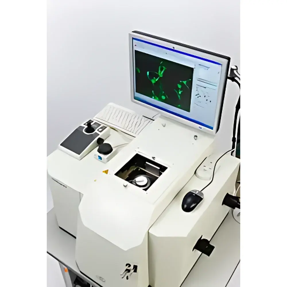
ECI TAXIScan-FL Fluorescent Cell Chemotaxis Dynamic Imaging System
| Origin | Japan |
|---|---|
| Manufacturer Type | Authorized Distributor |
| Import Status | Imported |
| Model | TAXIScan-FL |
| Price Range | USD 140,000 – 280,000 (FOB Japan) |
| Product Category | Live-Cell Chemotaxis Analysis System |
| Application Domain | Academic & Preclinical Research |
| Excitation Source | None (Requires External Fluorescence Illumination) |
| Detection Channels | 1 (Single-Channel Fluorescence + Brightfield) |
| Spatial Resolution | ≤30 µm |
| Fluorescence Sensitivity | ≥0.1 ng/mL (FITC-equivalent) |
| Imaging Throughput | Up to 12 Concurrent Assays |
| Sample Requirement | As Few as 100 Viable Cells per Assay |
| Temperature Control Range | RT + 3°C to 40°C |
| Microchannel Depths | 4, 5, 6, or 8 µm |
| Objective Lenses | 10×, 20×, 40×, 100× (Oil Immersion Compatible) |
| Filter Sets | Standard B/G/R Cube Configuration |
| Data Output Format | TIFF/AVI Time-Lapse Sequences with Metadata Embedding |
| Software Compliance | FDA 21 CFR Part 11–Ready Audit Trail, GLP-Compliant Experiment Logging |
Overview
The ECI TAXIScan-FL Fluorescent Cell Chemotaxis Dynamic Imaging System is a precision-engineered platform for quantitative, real-time analysis of live-cell chemotactic behavior under controlled chemical gradient conditions. Unlike conventional transwell or Boyden chamber assays, the TAXIScan-FL employs a proprietary silicon-based microfluidic chip architecture to generate stable, reproducible, and spatially defined linear or exponential chemokine gradients across planar microchannels—enabling direct visualization and statistical quantification of directional migration, morphodynamic remodeling, and fluorescence-labeled intracellular events (e.g., Ca2+ flux, ROS generation, granule exocytosis) at single-cell resolution. The system operates on the principle of laminar diffusion-controlled gradient formation within subcellular-height microchannels (4–8 µm depth), ensuring physical confinement of suspended cells in a quasi-2D imaging plane while maintaining physiological viability over extended observation periods (up to 24 h). Its optical design integrates an inverted cold-CCD imaging module with coaxial epi-illumination, high-NA objectives, and motorized autofocus—optimized for low-phototoxicity, long-term time-lapse acquisition without darkroom requirements.
Key Features
- Silicon microchip with 12 independent, parallel microchannels—each configurable with precise channel depths (4, 5, 6, or 8 µm) to match cell diameter and constrain motility in the XY plane
- Passive diffusion-based gradient generation enabling stable, non-perturbing chemokine concentration profiles (0–100 nM range typical) validated via computational fluid dynamics (CFD) modeling and fluorescent dye calibration
- Motorized, software-controlled autofocus system with sub-micron repeatability for continuous tracking of individual migrating cells across multi-hour experiments
- Integrated brightfield + single-channel fluorescence imaging (B/G/R filter sets) supporting common fluorophores including FITC, TRITC, and DAPI; no integrated laser source required—compatible with external LED or mercury lamp excitation
- Automated image acquisition and annotation: time-stamped AVI/TIFF sequences embedded with environmental metadata (temperature, timestamp, objective, channel ID)
- Onboard temperature regulation (RT + 3°C to 40°C) with ±0.3°C stability, ensuring consistent cellular metabolism and receptor kinetics during assay runtime
Sample Compatibility & Compliance
The TAXIScan-FL supports primary human and murine immune cells—including neutrophils, eosinophils, monocytes, T lymphocytes—as well as adherent and suspension-cultured lines (e.g., HL-60, THP-1, MDA-MB-231, U87MG, HUVEC, iPSC-derived neurons). Minimal sample input (≤100 viable cells per channel) reduces biological variability and enables rare-cell analysis (e.g., circulating tumor cells, tissue-resident macrophages). All hardware and firmware comply with IEC 61000-6-3 (EMC) and IEC 61010-1 (safety). Software architecture conforms to GLP and preclinical GMP documentation standards, including full audit trail logging, electronic signature support, and 21 CFR Part 11–compliant user access control. Experimental protocols align with ASTM E3094–20 (Standard Guide for Quantitative Chemotaxis Assay Validation) and ISO 20947–2 (Biotechnology — Cell Migration Assays — Performance Criteria).
Software & Data Management
The proprietary TAXIScan Analysis Suite provides end-to-end workflow automation—from gradient profile simulation and experimental parameter definition to post-acquisition segmentation, trajectory reconstruction, and statistical inference. Key modules include: Gradient Mapping Engine (quantifies spatial ligand distribution via calibration curve interpolation); CellTrack Pro (performs adaptive thresholding, centroid tracking, and velocity vector field computation); MorphoMetrics (extracts >30 shape descriptors: aspect ratio, circularity, protrusion frequency, leading edge dynamics); and Chemotactic Index Calculator (computes directionality bias relative to gradient axis using cosine-weighted angular distribution). All datasets export to HDF5 or CSV with MIAME-compliant metadata headers. Raw movie files are stored with embedded DICOM-SR–style structured reports for traceable data provenance.
Applications
- Chemotaxis Mechanistic Studies: Quantification of GPCR-mediated leukocyte navigation in inflammation models; evaluation of CXCR4/CXCL12, CCR5/CCL5, or FPR1/fMLP signaling axis perturbations
- Tumor Cell Invasion Modeling: Real-time monitoring of EMT-associated motility shifts in response to TGF-β, EGF, or stromal-derived factor gradients
- Drug Screening & Pharmacodynamics: High-content assessment of small-molecule inhibitors targeting PI3Kγ, ROCK, or WASp—measuring dose-dependent suppression of directional persistence
- Neuroimmune Crosstalk: Tracking microglial chemotaxis toward ATP or fractalkine gradients in co-culture with neuronal spheroids
- Allergic & Autoimmune Pathophysiology: Basophil and mast cell degranulation kinetics correlated with migration onset under IgE-antigen challenge
FAQ
What cell types are validated for use with the TAXIScan-FL system?
Primary human peripheral blood mononuclear cells (PBMCs), isolated neutrophils, murine bone marrow–derived macrophages, and over 27 adherent/suspension cell lines have been experimentally validated in peer-reviewed publications.
Is external fluorescence excitation required?
Yes—the system does not include an integrated light source. Users must supply compatible LED or arc-lamp illumination matched to their fluorophore’s excitation spectrum.
Can gradient steepness be modified during an ongoing experiment?
No—gradients are established passively prior to cell loading and remain stable for the duration of imaging; however, multiple chips with pre-configured depths and inlet geometries allow rapid method switching between gradient profiles.
Does the software support batch analysis of multi-channel experiments?
Yes—TAXIScan Analysis Suite processes all 12 channels in parallel, applying identical segmentation and tracking parameters, and exports consolidated statistics per condition in Excel-ready format.
What is the minimum viable cell count per assay?
As few as 50–100 morphologically intact, non-apoptotic cells yield statistically robust directional metrics when analyzed over ≥60 min time series.
Related Products