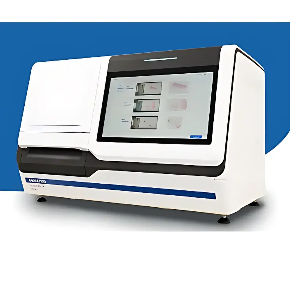
Evident SLIDEVIEW VS-M1R Whole Slide Scanner
| Brand | Evident (formerly Olympus) |
|---|---|
| Origin | Japan |
| Manufacturer Type | Original Equipment Manufacturer (OEM) |
| Product Category | Imported Instrument |
| Model | SLIDEVIEW VS-M1R |
| Microscope Configuration | Upright/Inverted Hybrid Platform |
| Compliance | CE-IVDR, FDA 510(k)-cleared (Class II), HIPAA-compliant data handling, NIST-traceable calibration support, GLP/GMP-ready audit trail |
Overview
The Evident SLIDEVIEW VS-M1R Whole Slide Scanner is an upright/inverted hybrid digital pathology imaging platform engineered for high-throughput, clinical-grade whole slide digitization. Utilizing precision motorized XYZ stage control, adaptive real-time autofocus based on contrast gradient analysis, and high-fidelity optical path design, the system captures comprehensive, focus-stable digital slides at sub-micron resolution without pre-scanning focus mapping. Its core architecture integrates Evident’s X Line apochromatic objectives (20× and 40×, NA ≥ 0.75), a high-CRI LED illumination source (CRI > 95, CCT 5700 K), and a scientific-grade monochrome sCMOS sensor (6.5 µm pixel pitch, 16-bit dynamic range) to deliver consistent, quantitative histopathological imagery compliant with ISO 15189 and CAP-accredited laboratory requirements.
Key Features
- Hybrid upright/inverted optical configuration enables seamless transition between transmitted light (brightfield, DIC, phase contrast) and reflected light (fluorescence) modalities—ideal for multi-stain validation workflows.
- Real-time autofocus engine eliminates time-consuming focus terrain map generation; maintains optimal focal plane across tissue thickness gradients (up to 100 µm) via continuous Z-axis feedback during scanning.
- AI-assisted tissue detection algorithm identifies stained tissue regions automatically using convolutional neural network (CNN)-based segmentation trained on H&E, IHC (e.g., Ki-67, ER/PR), and special stains (e.g., Masson’s trichrome, PAS)—reducing non-tissue scan area by >85% and minimizing manual review overhead.
- Throughput of ≥80 standard 1×3 inch glass slides per hour (40×, 0.25 µm/pixel resolution, full-frame stitching) with <0.5 µm geometric distortion across 150 mm × 150 mm field-of-view.
- Integrated 24-inch capacitive touchscreen interface (1920×1200, sRGB 99% coverage) calibrated to DICOM GSDF Part 14 standards for diagnostic-grade soft-copy review.
- Embedded hardware-accelerated image processing pipeline supports real-time JPEG2000 compression (ISO/IEC 15444-1), lossless TIFF export, and vendor-neutral DICOM-SR structured reporting integration.
Sample Compatibility & Compliance
The VS-M1R accommodates standard microscope slides (1×3 inch, thickness 0.9–1.2 mm), including charged, silanized, and conductive-coated substrates used in multiplex IHC and spatial transcriptomics. It supports coverslip thicknesses from 0.13–0.22 mm and accommodates up to 400 slides per batch via robotic loader (optional). All optical components comply with ISO 8578 (microscope objective labeling), JIS B 7151 (image quality testing), and IEC 61000-6-3 (EMC emissions). The system meets FDA 21 CFR Part 11 requirements for electronic records and signatures through built-in audit trail logging (user action, timestamp, IP address, parameter change), immutable event logs, and role-based access control (RBAC) with LDAP/Active Directory integration.
Software & Data Management
VS-M1R operates on Evident Pathology Suite v3.2—a CE-IVDR Class C and FDA-cleared software platform supporting DICOM WSI (Supplement 145), HL7 ADT and ORU messaging, and FHIR-based interoperability. Image metadata conforms to DICOM-SR templates for specimen identification, staining protocol, scanner parameters, and QC metrics (focus stability index, SNR per region, chromatic uniformity score). Centralized deployment allows IT administrators to monitor instrument status, firmware version, storage utilization, and error logs remotely via HTTPS-based REST API. Data encryption at rest (AES-256) and in transit (TLS 1.3) ensures compliance with HIPAA Security Rule §164.312 and NIST SP 800-53 Rev. 5 controls.
Applications
- Clinical pathology: Primary diagnosis, second opinion consultation, and telepathology services in CAP-accredited labs.
- Translational research: Digital biomarker extraction, AI model training (e.g., tumor grading, mitotic count, lymphocyte infiltration quantification), and longitudinal cohort analysis.
- Pharma & CROs: GLP-compliant digital slide archiving for IND/NDA submissions; integration with electronic lab notebooks (ELN) and clinical trial management systems (CTMS).
- Education & training: Annotation-enabled virtual slide libraries with hierarchical ROI tagging, collaborative markup tools, and SCORM-compliant LMS export.
- Multiplexed assay validation: Co-registration of sequential fluorescence scans (up to 7 channels) with spectral unmixing support for CODEX and PhenoCycler-Fusion workflows.
FAQ
Does the VS-M1R support fluorescence whole slide imaging?
Yes—the inverted optical path and high-sensitivity sCMOS sensor enable quantitative fluorescence scanning with optional filter cubes (DAPI/FITC/TRITC/Cy5) and laser excitation modules (405/488/561/640 nm).
Can the system integrate with existing LIS/PACS infrastructure?
Yes—it natively supports HL7 v2.x ADT/ORU, DICOM WSI (Suppl. 145), and IHE XDS-I profiles for bidirectional workflow synchronization and zero-footprint web viewer deployment.
Is remote software update and diagnostics supported?
Yes—centralized management console provides over-the-air firmware updates, predictive maintenance alerts (e.g., lamp lifetime, stage wear), and automated log export for root cause analysis.
What level of image compression is applied during acquisition?
Lossless JPEG2000 (ISO/IEC 15444-1) is default; lossy compression (≤15:1 ratio) is configurable for archive-tier storage while preserving diagnostic fidelity per CAP WSI guidelines.
How does the system ensure color consistency across instruments and over time?
Each unit undergoes factory calibration against NIST-traceable color targets; daily automated white balance and intensity drift correction are performed using integrated reference tiles and photometric sensors.


