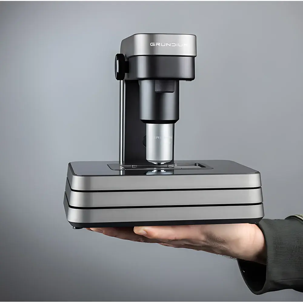
Grundium Ocus 20 Digital Pathology Slide Scanner
| Brand | Grundium |
|---|---|
| Origin | Finland |
| Model | Ocus 20 |
| Dimensions (W×D×H) | 18 cm × 18 cm × 19 cm |
| Weight | 3.5 kg |
| Objective Resolution (20×) | 0.48 µm/pixel |
| Wide-field Lens Resolution (1×) | 10 µm/pixel |
| Scanning Area | Up to 15 mm × 15 mm per field |
| Scan Time (typical) | ~2 min per slide |
| Autofocus | Z-axis multi-layer (up to 5 layers), per-field dynamic focus during continuous motion |
| Stage | Motorized XY stage, compatible with standard 75 mm × 25 mm glass slides |
| Storage | Internal 500 GB SSD, expandable via USB |
| Connectivity | 1 GbE Ethernet, IEEE 802.11ac Wi-Fi, hotspot mode |
| Image Formats | TIFF, SVS, MRXS |
| Sensor | 6 MP monochrome CMOS |
| Illumination | Köhler-optimized LED |
| User Interface | Web-based, browser-native (Chrome, Edge, Safari, Firefox), touch-enabled |
| Onboard Processing | Dual-core embedded processor, real-time rendering and stitching |
| Software Features | Region-of-interest (ROI) selection (freehand/polygonal), tile-based stitching, Z-stack export, GLP-compliant metadata logging |
Overview
The Grundium Ocus 20 Digital Pathology Slide Scanner is a compact, high-fidelity whole-slide imaging (WSI) system engineered for precision, reproducibility, and workflow integration in clinical pathology, translational research, and veterinary diagnostics. Utilizing a Köhler-optimized LED illumination path and a calibrated 6-megapixel monochrome CMOS sensor, the Ocus 20 captures high-contrast, color-accurate digital slides at native optical resolution—0.48 µm/pixel with the 20× objective and 10 µm/pixel using the 1× macro lens. Its core imaging architecture relies on continuous-motion scanning combined with per-field dynamic autofocus, eliminating focus drift across heterogeneous tissue topographies. Unlike legacy step-and-repeat scanners, the Ocus 20 acquires each field independently in real time, adjusting Z-position before exposure—a critical capability for frozen sections, thick cytology preparations, or unevenly mounted specimens. The integrated dual-core processing unit handles image reconstruction, lossless compression, and seamless tile-based stitching without external compute dependency.
Key Features
- Real-time, per-field autofocus during continuous XY scanning—ensures optimal focus across variable tissue thicknesses and coverslip heights.
- Z-axis multi-layer acquisition (up to 5 focal planes per field), supporting extended-depth-of-field (EDF) reconstruction and quantitative Z-stack analysis.
- Web-native control interface: no proprietary software installation required; compatible with Chrome, Edge, Safari, and Firefox on Windows, macOS, iOS, Android, and Linux.
- Compact footprint (18 × 18 × 19 cm) and lightweight design (3.5 kg) enable deployment in space-constrained environments—including operating rooms, field clinics, and mobile diagnostic units.
- Onboard 500 GB SSD with USB 3.0 expansion support ensures secure, local storage of DICOM-compliant TIFF, Aperio SVS, and MRXS files—fully traceable with embedded EXIF and DICOM-SR metadata.
- Motorized XY stage precisely accommodates standard 75 mm × 25 mm glass slides; ROI selection supports freehand, polygonal, and rectangular annotation prior to scan initiation.
Sample Compatibility & Compliance
The Ocus 20 supports a broad spectrum of specimen types across human, veterinary, botanical, and materials science domains. It routinely digitizes H&E-stained formalin-fixed paraffin-embedded (FFPE) sections, rapid-frozen intraoperative specimens, liquid-based cytology smears (e.g., Pap, FNA), and Mohs micrographic surgery margins. In veterinary practice, it enables remote review of canine lymphoma biopsies or equine dermatopathology slides. Plant histology (e.g., vascular bundle cross-sections) and microbiological preparations (Gram-stained smears, fungal mounts) are imaged with consistent contrast and spatial fidelity. For biomedical materials, the system validates fiber morphology in wool or cashmere quality control and assesses scaffold porosity in tissue-engineered constructs. All image acquisitions comply with ISO 15189 pre-analytical documentation requirements and support audit-ready metadata fields aligned with CLIA and CAP checklist items. Optional DICOM-SR export facilitates PACS integration under IHE XDS-I profiles.
Software & Data Management
The embedded web application provides full control over acquisition parameters—including exposure time, white balance, ROI definition, Z-stack depth, and compression level—via intuitive sliders and toggles. Each scanned slide is automatically tagged with timestamp, operator ID, microscope objective used, and environmental metadata (e.g., ambient temperature logged via onboard sensor). Image files include embedded ICC profiles and calibration grids compliant with ASTM E2912-13 (Standard Practice for Digital Microscopy Calibration). The system supports FDA 21 CFR Part 11–ready user authentication when deployed behind institutional identity providers (LDAP/SAML), and maintains immutable audit trails for all acquisition and annotation events. Export workflows integrate natively with QIAGEN Digital Insights, Philips IntelliSite, and 3DHISTECH platforms via DICOM WADO-URI or RESTful APIs.
Applications
- Clinical pathology: Primary diagnosis, second-opinion consultation, and telepathology triage—especially in resource-limited or rural settings.
- Intraoperative frozen section analysis: Rapid digitization (<2 min/slide) enables real-time expert review during Mohs surgery or tumor margin assessment.
- Education and training: High-resolution WSI libraries support competency-based assessment of trainee pattern recognition across hematology, dermatopathology, and neuropathology curricula.
- Preclinical research: Longitudinal tracking of murine tumor xenograft histomorphology and immune cell infiltration quantification using open-source tools (QuPath, HALO).
- Veterinary diagnostics: Remote case review between field practitioners and board-certified pathologists—validated for canine mast cell tumors and feline chronic kidney disease staging.
- Biotech QA/QC: Verification of single-cell chip uniformity, hydrogel degradation kinetics, and electrospun nanofiber alignment in regenerative medicine R&D.
FAQ
Does the Ocus 20 require a dedicated workstation or server to operate?
No. All image capture, processing, stitching, and storage occur onboard. A standard web browser on any network-connected device serves as the sole interface.
Can the system be integrated into an existing LIS/PACS environment?
Yes—via DICOM WADO-URI, REST API, or direct file ingestion (SVS/TIFF) with configurable metadata mapping to HL7 ADT or IHE XDS-I standards.
Is Z-stack data export supported for 3D reconstruction?
Yes. Per-field Z-series (up to 5 layers) are exported as multi-page TIFF or OME-TIFF with precise Z-interval metadata, compatible with Imaris, Bitplane, and Fiji.
What regulatory certifications does the Ocus 20 hold?
CE-marked as a Class I medical device under MDR 2017/745; FDA 510(k) clearance pending; conforms to IEC 61000-6-3 (EMC) and IEC 61000-6-1 (immunity).
How is data security managed during wireless transmission?
All Wi-Fi communications use WPA3-Enterprise encryption; hotspot mode enforces TLS 1.3 for browser sessions; locally stored images are AES-256 encrypted at rest.
Related Products


