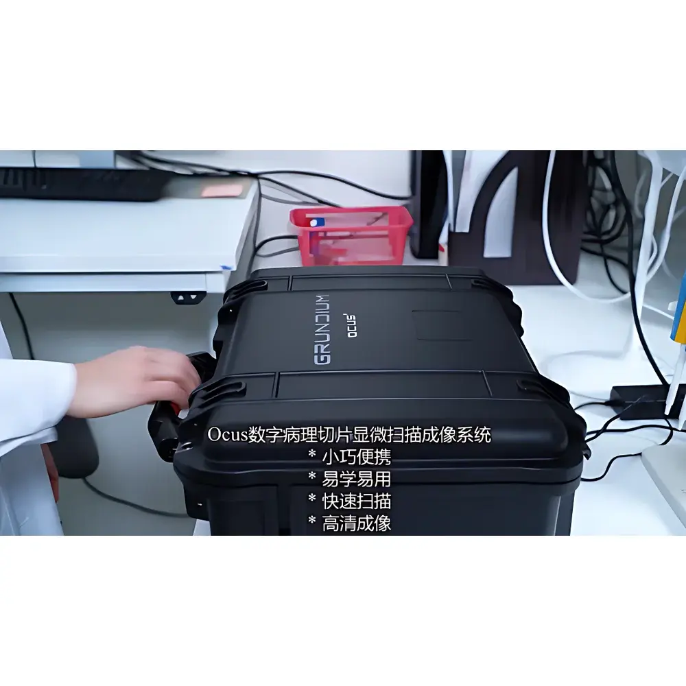
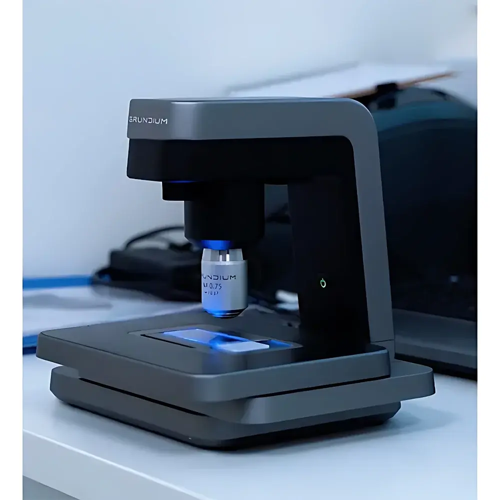
Grundium Ocus 40 Digital Pathology Slide Scanner
| Brand | Grundium |
|---|---|
| Origin | Finland |
| Model | Ocus 40 |
| Imaging Resolution (40× objective) | 0.25 µm/pixel |
| Wide-field Lens Resolution (1×) | 10 µm/pixel |
| Scan Area | Up to 15 mm × 15 mm per region |
| Scan Time (typical) | ~3 min per 15×15 mm region |
| Z-stack Capability | Up to 5 focal planes |
| Storage | Internal 500 GB SSD, expandable via USB |
| Connectivity | 1 GbE Ethernet, IEEE 802.11ac Wi-Fi, hotspot mode |
| Image Formats | TIFF, SVS, MRXS |
| Dimensions (W×D×H) | 18 cm × 18 cm × 19 cm |
| Weight | 3.5 kg |
| Illumination | Köhler-optimized LED |
| Sensor | 6 MP monochrome CMOS |
| Stage | Motorized XY stage compatible with standard 75 mm × 25 mm glass slides |
| Focus | Real-time dynamic autofocus per field-of-view during continuous scanning |
| Software Interface | Browser-based (HTML5), touch-enabled, no client installation required |
| OS Compatibility | Windows, macOS, iOS, Android |
Overview
The Grundium Ocus 40 is a compact, high-performance digital pathology slide scanner engineered for precision, reproducibility, and workflow flexibility in clinical, research, and educational environments. Unlike traditional slide scanners reliant on external workstations or proprietary software ecosystems, the Ocus 40 integrates a dual-core embedded processing unit and dedicated NVIDIA graphics acceleration directly into the instrument—enabling full image acquisition, real-time autofocus, tile stitching, and format conversion (TIFF/SVS/MRXS) without external computing dependencies. Its optical architecture employs Köhler illumination with a stable, long-life LED source to ensure uniform intensity and minimal photobleaching across extended scanning sessions. The system operates on a continuous-motion scanning principle, where the motorized XY stage moves at controlled velocity while the camera captures images at precisely timed intervals—each frame independently autofocused in Z using contrast-based algorithms optimized for histological contrast. This eliminates focus drift artifacts common in step-and-repeat systems and ensures consistent sharpness across heterogeneous tissue regions, including those with variable thickness or refractive index (e.g., frozen sections, cytology smears, or decalcified bone).
Key Features
- Integrated dual-processor architecture with onboard NVIDIA GPU for real-time image processing, eliminating dependency on external PCs or servers.
- Browser-based HTML5 user interface—accessible from any modern web browser on Windows, macOS, iOS, or Android devices; no app installation or driver configuration required.
- Dynamic per-field autofocus during continuous scanning—each field-of-view receives individual Z-height optimization before capture, ensuring optimal resolution across morphologically complex specimens.
- Z-stack acquisition support for up to five focal planes per location, enabling extended-depth-of-field reconstruction or 3D tissue analysis workflows.
- Flexible region selection: users may define multiple irregular ROIs on a single slide via intuitive touch-enabled interface; only selected areas are scanned, reducing storage overhead and processing time.
- Compact footprint (18 × 18 × 19 cm) and lightweight design (3.5 kg) facilitate deployment in space-constrained settings—including operating rooms, field clinics, teaching labs, and mobile diagnostic units.
- Native support for industry-standard digital pathology formats: uncompressed TIFF for archival integrity, Aperio SVS for PACS integration, and MRXS for multi-resolution pyramid delivery.
Sample Compatibility & Compliance
The Ocus 40 accommodates standard 75 mm × 25 mm glass microscope slides, supporting routine H&E-stained sections, IHC/IF preparations, cytology smears, fine-needle aspirates, and frozen-section specimens. Its optical path is calibrated for both brightfield and phase contrast imaging modalities, making it suitable for unstained or lightly stained samples such as microbiological smears or plant tissue sections. The system meets essential requirements for GLP-compliant documentation: audit-trail logging of all acquisition parameters (objective used, exposure time, Z-position, ROI coordinates), timestamped metadata embedding, and immutable image file generation. While not FDA-cleared as a primary diagnostic device, its output conforms to DICOM Supplement 145 (Whole Slide Imaging) standards and supports integration into ISO 15189-accredited laboratory information systems. For regulated environments, optional secure network configuration (TLS 1.2+ encryption, role-based access control) aligns with HIPAA and GDPR data handling expectations.
Software & Data Management
All image acquisition, annotation, and export functions are executed through a responsive web application served directly from the scanner’s internal Linux OS. No local software installation is required—users interact via Chrome, Edge, Safari, or Firefox on desktops, tablets, or smartphones. The built-in image browser supports lossless zoom, panning, color balance adjustment, and ROI measurement (area, perimeter, pixel count). Export options include full-resolution TIFF stacks, pyramidal SVS files compatible with OpenSlide libraries, and MRXS containers for cloud-based viewing platforms. Local storage consists of a 500 GB SSD; additional capacity can be added via USB 3.0–connected drives. Network transfer supports DICOM WADO-URI, SFTP, and HTTP POST endpoints, enabling automated ingestion into PACS, LIS, or LIMS infrastructures. All metadata—including scanner serial number, operator ID (if authenticated), date/time stamps, and objective magnification—is embedded in EXIF and Aperio-compatible headers.
Applications
The Ocus 40 serves diverse use cases across human and veterinary pathology, biomedical research, and quality assurance. In clinical pathology, it supports rapid intraoperative consultation (e.g., Mohs surgery margin assessment), remote second-opinion review, and telepathology initiatives in underserved regions. In academic settings, it enables scalable digitization of teaching slide collections and facilitates collaborative annotation projects. Veterinary and agricultural laboratories utilize it for post-mortem diagnostics and parasite identification in animal tissue or fecal smears. Plant biologists apply it to leaf cross-sections and root nodule imaging, while materials scientists employ its high-resolution capability for fiber morphology analysis (e.g., cashmere grading) or biomaterial scaffold characterization. Additionally, the system is deployed in QC workflows for microfluidic chip fabrication, where single-cell array integrity must be verified prior to sequencing.
FAQ
Does the Ocus 40 require a dedicated computer or server to operate?
No—the scanner contains an embedded industrial-grade computer with dual processors and integrated GPU. All imaging, stitching, and export functions run locally.
Can the system scan unstained or phase-contrast specimens?
Yes—its Köhler LED illumination and high-sensitivity 6 MP monochrome sensor support low-contrast specimens without staining.
Is the software compatible with hospital PACS or LIS systems?
Yes—SVS and MRXS outputs are compatible with major PACS vendors; DICOM WADO-URI and SFTP interfaces enable direct integration.
What level of cybersecurity does the device support?
It supports TLS 1.2+ encrypted web access, configurable user authentication, and audit-log export for compliance with HIPAA, GDPR, and ISO 27001 frameworks.
How is focus maintained across uneven or thick tissue sections?
Each field-of-view undergoes independent Z-axis autofocus immediately before image capture, compensating for local topography variations in real time.
Related Products


