Hamamatsu PDE Fluorescence Image-Guided Surgical Navigation System
| [Brand | Hamamatsu |
|---|---|
| Origin | Japan |
| Manufacturer | Hamamatsu Photonics K.K. |
| Type | Import |
| Model | PDE |
| Power Supply | AC 100 V ±10 V, 50/60 Hz |
| Max Power Consumption | 50 W |
| Operating Temperature | +10 °C to +40 °C |
| Storage Temperature | +10 °C to +40 °C |
| Operating Humidity | 20–70 % RH (non-condensing) |
| Storage Humidity | 20–70 % RH (non-condensing) |
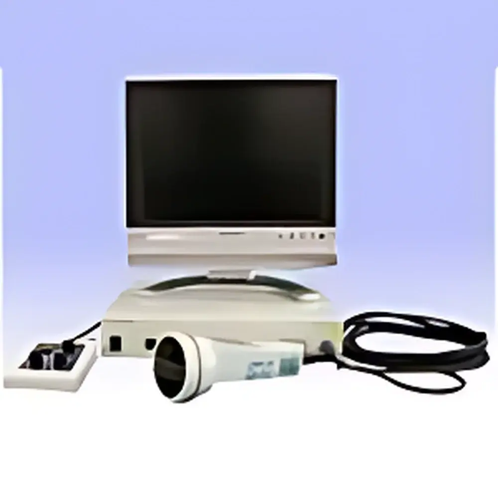
| Camera Unit Dimensions (W×D×H) | ≈80 mm × 181 mm × 80 mm (excl. protrusions), Mass: ≈0.5 kg (excl. cable) |
| Controller Unit Dimensions (W×D×H) | ≈322 mm × 283 mm × 55 mm (excl. protrusions), Mass: ≈2.8 kg |
| Video Output | PAL-standard analog video signal (composite) |
| Detection Band | Near-infrared (NIR), centered at ~800–850 nm for ICG fluorescence] |
Overview
The Hamamatsu PDE Fluorescence Image-Guided Surgical Navigation System is a compact, CE-marked, clinically validated NIR fluorescence imaging platform engineered for real-time intraoperative visualization of indocyanine green (ICG)-labeled anatomical structures. It operates on the principle of near-infrared fluorescence (NIRF) imaging: an integrated 785 nm or 805 nm excitation source illuminates tissue; ICG—administered via subcutaneous, intradermal, or intravascular injection—absorbs this light and re-emits photons at ~830–850 nm. The system’s high-sensitivity InGaAs or silicon-based NIR camera detects this emitted fluorescence through spectral filtering, enabling real-time, non-invasive, and label-specific visualization of lymphatic vessels, sentinel lymph nodes, perfused vasculature, and tissue viability margins. Unlike visible-light or white-light imaging, the PDE leverages the optical transparency window of biological tissue (700–900 nm), where hemoglobin and water absorption are minimized—permitting photon penetration up to 10–15 mm in adipose tissue and ~5–8 mm in muscle-dense regions. This physical basis underpins its clinical utility across oncologic, cardiovascular, and reconstructive surgical disciplines.
Key Features
- Real-time dynamic fluorescence imaging with sub-second latency and continuous analog video output (PAL standard)
- Integrated excitation source and narrowband NIR detection optics optimized for ICG’s excitation/emission profile
- Compact, modular architecture: lightweight handheld camera unit (0.5 kg) and dedicated controller console (2.8 kg) designed for sterile field integration
- No external PC required for basic operation; analog video output compatible with standard medical monitors, endoscopy towers, and DICOM-compliant video capture systems
- Optimized ambient light rejection: system performance validated under operating room lighting conditions; halogen/xenon surgical lights must be dimmed or switched off during acquisition due to spectral overlap in the 750–850 nm band
- Robust thermal management ensuring stable operation across 10–40 °C ambient range and ≤70 % non-condensing humidity
Sample Compatibility & Compliance
The PDE is intended for use with FDA-cleared and CE-marked ICG formulations (e.g., Akorn, Diagnostic Green) administered per approved dosing protocols (typically 0.2–0.5 mg/kg). It does not require radioactive tracers or ionizing radiation. While ICG is the primary fluorophore, the system’s spectral response permits limited visualization of endogenous chromophores—including deoxygenated hemoglobin—in superficial vasculature without exogenous contrast, though signal-to-noise ratio remains significantly lower than ICG-enhanced imaging. The device complies with IEC 60601-1 (medical electrical equipment safety), IEC 62304 (software lifecycle), and IEC 60601-2-57 (particular requirements for optical equipment). It supports GLP-aligned documentation workflows when paired with validated video capture hardware and timestamped storage solutions compliant with 21 CFR Part 11 audit trail requirements.
Software & Data Management
The PDE outputs uncompressed PAL-format composite video—a deterministic, low-latency analog interface that ensures compatibility with legacy OR infrastructure and avoids digital compression artifacts. For digital archiving, integration with third-party video capture cards (e.g., Epiphan, DVI2PCIe Duo) enables frame-accurate recording into PACS or EMR-adjacent repositories. Metadata tagging (e.g., procedure type, time stamp, operator ID) requires external middleware or custom DICOM-SR wrappers. No proprietary software is embedded in the core unit; firmware updates are performed via USB using Hamamatsu-provided utilities under controlled change management per ISO 13485. Exported video files retain native resolution (720 × 576 pixels) and are suitable for retrospective surgical workflow analysis, peer review, and regulatory submission dossiers.
Applications
- Sentinel lymph node mapping in breast cancer, melanoma, and gynecologic malignancies—guiding minimally invasive biopsy and reducing unnecessary axillary dissection
- Intraoperative assessment of graft patency and anastomotic flow in coronary artery bypass grafting (CABG) and peripheral bypass procedures
- Real-time evaluation of flap perfusion and vascular compromise in microsurgical reconstruction (e.g., DIEP, ALT flaps)
- Visualization of biliary anatomy during cholecystectomy and hepatic resection to prevent ductal injury
- Neurovascular mapping in cerebrovascular surgery—identifying cortical surface vessels and assessing collateral flow
- Preclinical and translational research in small-animal models of tumor metastasis, lymphatic dysfunction, and ischemia-reperfusion injury
FAQ
What is the maximum tissue depth at which ICG fluorescence can be reliably detected?
In subcutaneous adipose tissue, the PDE resolves ICG fluorescence at depths up to 10–12 mm. In skeletal muscle or highly vascularized parenchyma, effective detection depth reduces to 5–7 mm due to increased scattering and hemoglobin absorption.
Can the PDE visualize blood vessels without ICG administration?
Yes—superficial veins (<2 mm depth) may generate detectable NIR signal due to hemoglobin absorption contrast, but sensitivity and specificity are markedly inferior to ICG-enhanced imaging. Clinical decision-making should rely exclusively on contrast-enhanced visualization.
Is the system compatible with sterile draping protocols?
The camera probe is designed for use with FDA-cleared sterile transparent sleeves (e.g., Medline, SteriShield); the controller unit remains outside the sterile field and connects via shielded coaxial cable.
Does the PDE support quantitative fluorescence intensity measurement?
No—the system delivers qualitative, real-time visual guidance only. Absolute radiometric calibration is not implemented; intensity comparisons across sessions require standardized illumination, distance, and exposure settings.
What regulatory clearances does the PDE hold?
The PDE is CE-marked under MDR 2017/745 Class IIa for image-guided surgical navigation. It is not currently 510(k)-cleared by the U.S. FDA; U.S. use is restricted to investigational or humanitarian device exemption (HDE) pathways pending institutional IRB approval.


