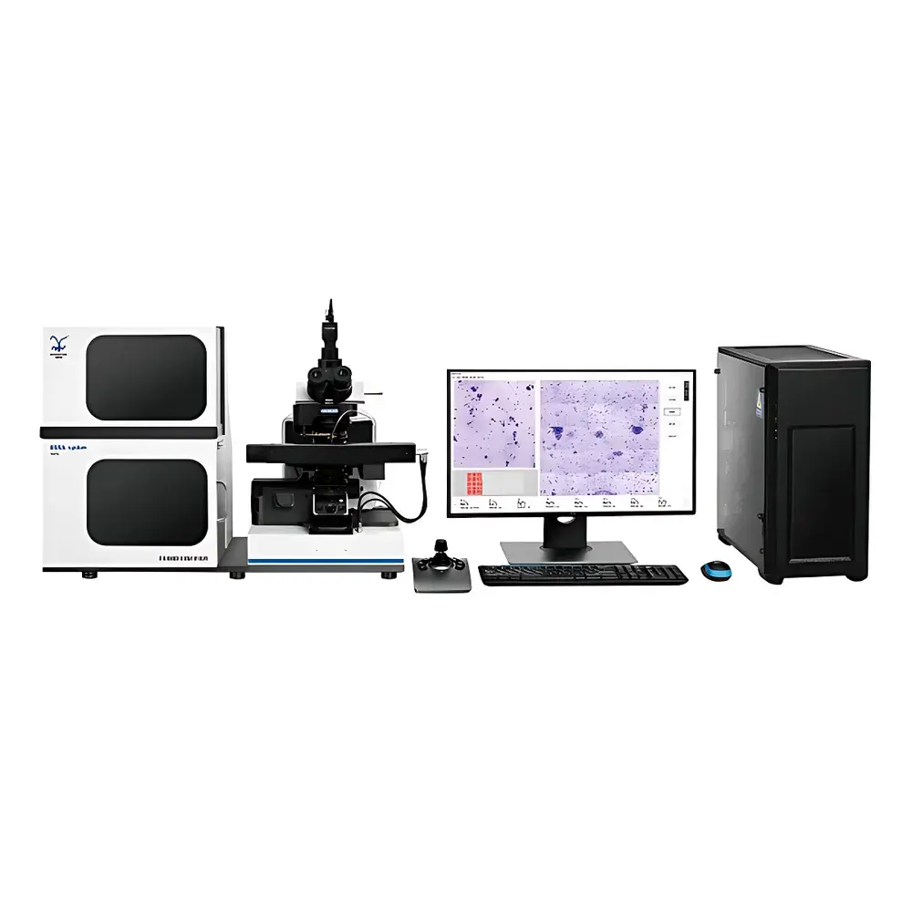
HRH-200A Automated Micronucleus and Chromosomal Aberration Image Analysis System
| Brand | Huironghe |
|---|---|
| Origin | Beijing, China |
| Model | HRH-200A |
| Type | Fully Automated Microscopic Image Analysis System for Cytogenetic Toxicology |
| Slide Capacity | ≥200 slides (batch loading), ≥8 slides on stage simultaneously |
| Objective Lenses | 10×, 20×, 100× (oil-immersion auto-dipping) |
| Analysis Modes | Real-time concurrent scanning & analysis |
| Cell Types Supported | Binucleated cells, mononucleated cells, polychromatic erythrocytes (PCE) |
| Output Metrics | Micronucleus frequency (%), micronucleated cell frequency (%), aberration classification (dicentrics, rings, fragments), audit-trail-enabled report export |
Overview
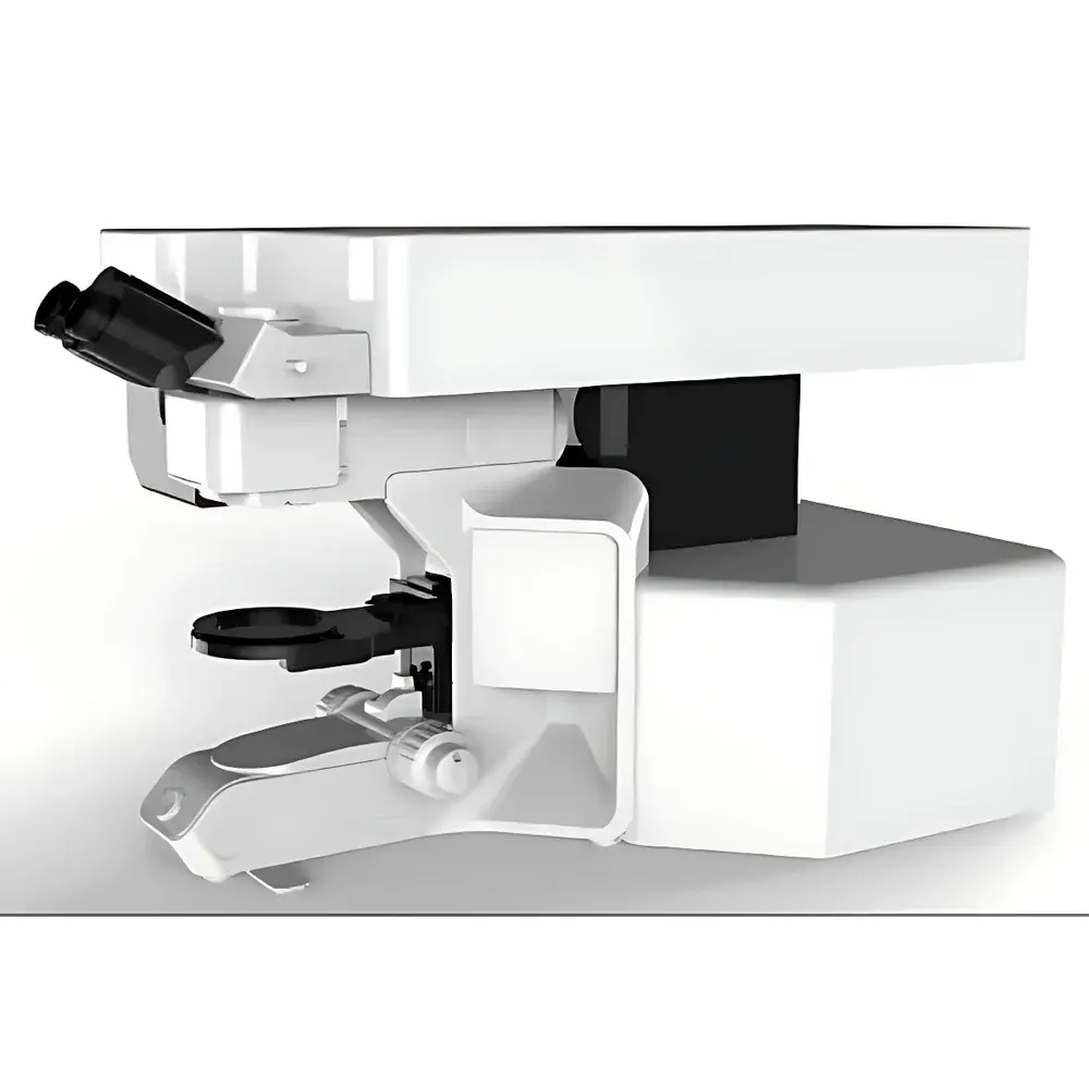
The HRH-200A Automated Micronucleus and Chromosomal Aberration Image Analysis System is a CE-compliant, laboratory-grade cytogenetic imaging platform engineered for high-throughput, standardized scoring of micronuclei (MN) and structural chromosomal aberrations in metaphase spreads and interphase nuclei. Designed in accordance with OECD Test Guideline 487 (In Vitro Mammalian Cell Micronucleus Test) and ICH S2(R2), the system integrates hardware automation with rule-based computer vision algorithms to replace manual microscopic enumeration—reducing intra- and inter-operator variability while maintaining full traceability under GLP conditions. Its core architecture employs brightfield and phase-contrast digital microscopy coupled with adaptive focus mapping, dynamic Z-stack acquisition, and multi-scale object detection to distinguish micronuclei (≥1/3 to ≤1/2 the diameter of the main nucleus, non-refractile, same staining intensity, located within the cytoplasm) from artifacts, nuclear buds, or apoptotic bodies. The system operates as a closed-loop workflow: from unattended slide loading through automated objective switching, oil-immersion lens deployment, metaphase-finding via texture-based pattern recognition, to pixel-level segmentation and morphometric validation of candidate micronuclei.
Key Features
- Fully automated slide handling: Robust robotic loader accommodates ≥200 standard 25 × 75 mm glass slides per batch; motorized stage holds ≥8 slides concurrently with barcode-scanning capability for sample tracking.
- Multi-objective scanning engine: Integrated 10× (overview scan), 20× (preliminary screening), and 100× oil-immersion objectives with automatic oil dispensing, meniscus control, and precision Z-axis stepping (±0.1 µm resolution).
- Real-time concurrent analysis: Image acquisition and AI-assisted micronucleus detection occur in parallel—no post-scan processing delay; results update dynamically during scanning.
- Cell-type–agnostic detection logic: Validated for binucleated lymphocytes (cytokinesis-block method), mononucleated cells, and rodent bone marrow polychromatic erythrocytes (PCEs), with configurable nuclear morphology thresholds.
- Interactive verification interface: Analysts review flagged candidates using side-by-side original image, segmentation mask, and feature overlay (area, circularity, intensity ratio, distance from main nucleus); corrections are logged with timestamp and operator ID.
- Regulatory-ready reporting: Generates PDF/Excel reports compliant with ISO/IEC 17025 documentation requirements, including raw image thumbnails, cell counts, MN frequencies, coefficient of variation (CV), and full audit trail per ISO 21 CFR Part 11 Annex 11 principles.
Sample Compatibility & Compliance
The HRH-200A accepts conventionally Giemsa-, DAPI-, or acridine orange-stained cytospin preparations and metaphase spreads on standard microscope slides. It supports both human peripheral blood lymphocyte cultures (with cytochalasin-B block) and rodent bone marrow smears. All image acquisition parameters—including exposure time, white balance, and gamma correction—are locked per protocol and stored with metadata. The system meets essential requirements of ISO 13485:2016 (medical device QMS), aligns with ASTM E2904-21 (standard guide for digital image analysis in genetic toxicology), and facilitates compliance with EU CLP Regulation Annex VI reporting formats. Validation packages include system suitability tests (SST) using reference slides with certified MN frequencies (e.g., NIST SRM 2366).
Software & Data Management
The embedded HRH-ImageAnalyzer v4.x software runs on Windows 10 IoT Enterprise LTSB with FIPS 140-2–validated cryptographic modules. It implements role-based access control (RBAC), electronic signatures, and immutable audit logs covering every user action—from slide registration to final report sign-off. Raw TIFF sequences (16-bit depth, lossless compression) are archived in hierarchical folder structures compliant with 21 CFR Part 11 §11.10(d). Data export supports DICOM-SR (Structured Reporting) for integration into LIS/HIS environments and CSV/JSON for statistical analysis in R or Python. Optional cloud backup adheres to GDPR Article 32 technical safeguards, with AES-256 encryption in transit and at rest.
Applications
- Genotoxicity assessment of food additives, pharmaceutical excipients, and cosmetic ingredients per OECD TG 487 and EFSA guidance.
- Biodosimetry triage following radiological incidents: rapid quantification of dicentric chromosomes and ring formations in peripheral blood lymphocytes.
- Occupational health surveillance of radiation workers under IAEA Safety Standards Series No. GSR Part 3.
- Preclinical safety pharmacology studies requiring chromosomal integrity endpoints in regulatory submissions (FDA IND, EMA IMPD).
- Academic research in DNA repair mechanisms, clastogen vs. aneugen classification, and dose-response modeling of mutagenic agents.
FAQ
Does the HRH-200A support fluorescence-stained preparations?
Yes—optional LED-based epi-fluorescence illumination module (365 nm/450–490 nm/515–560 nm excitation bands) enables DAPI, FITC, and Cy3-compatible protocols.
Can the system be validated for GLP-compliant studies?
Yes—comprehensive IQ/OQ/PQ documentation templates, calibration certificates for motorized components, and software validation scripts (including boundary testing and forced-error recovery) are provided.
What is the typical throughput for human lymphocyte samples?
Approximately 12–15 fully analyzed slides per hour (including metaphase finding, 100× imaging of ≥1000 binucleated cells, and MN scoring), depending on slide quality and aberration load.
Is remote diagnostics supported?
Yes—secure TLS 1.3–encrypted remote desktop access (with customer consent) enables firmware updates and troubleshooting without on-site service visits.
How are false positives minimized during micronucleus detection?
Through cascaded filtering: (1) intensity homogeneity thresholding, (2) spatial proximity constraint (micronucleus must reside within cytoplasmic boundary), (3) size–intensity correlation model trained on >50,000 expert-annotated objects, and (4) optional deep learning refinement layer (U-Net architecture) available as add-on module.