Invitrogen EVOS FLoid™ Cell Imaging Station
| Brand | Invitrogen |
|---|---|
| Origin | USA |
| Manufacturer | Thermo Fisher Scientific |
| Product Category | Imported |
| Model | FLoid |
| Pricing | Upon Request |
| Dimensions | 40.4 cm (W) × 53.6 cm (H) × 35.3 cm (D) |
| Weight | 11.8 kg |
| Power Input | 100–240 VAC, 50–60 Hz |
| DC Output | 5 V DC, 4.15 A |
| Operating Temperature | 4–32 °C |
| Operating Humidity | <90% RH (non-condensing) |
| Camera Sensor | Sony ICX445 EXview HAD CCD, 1.3 MP, 1/3″ format |
| Image Resolution | 1296 × 964 pixels |
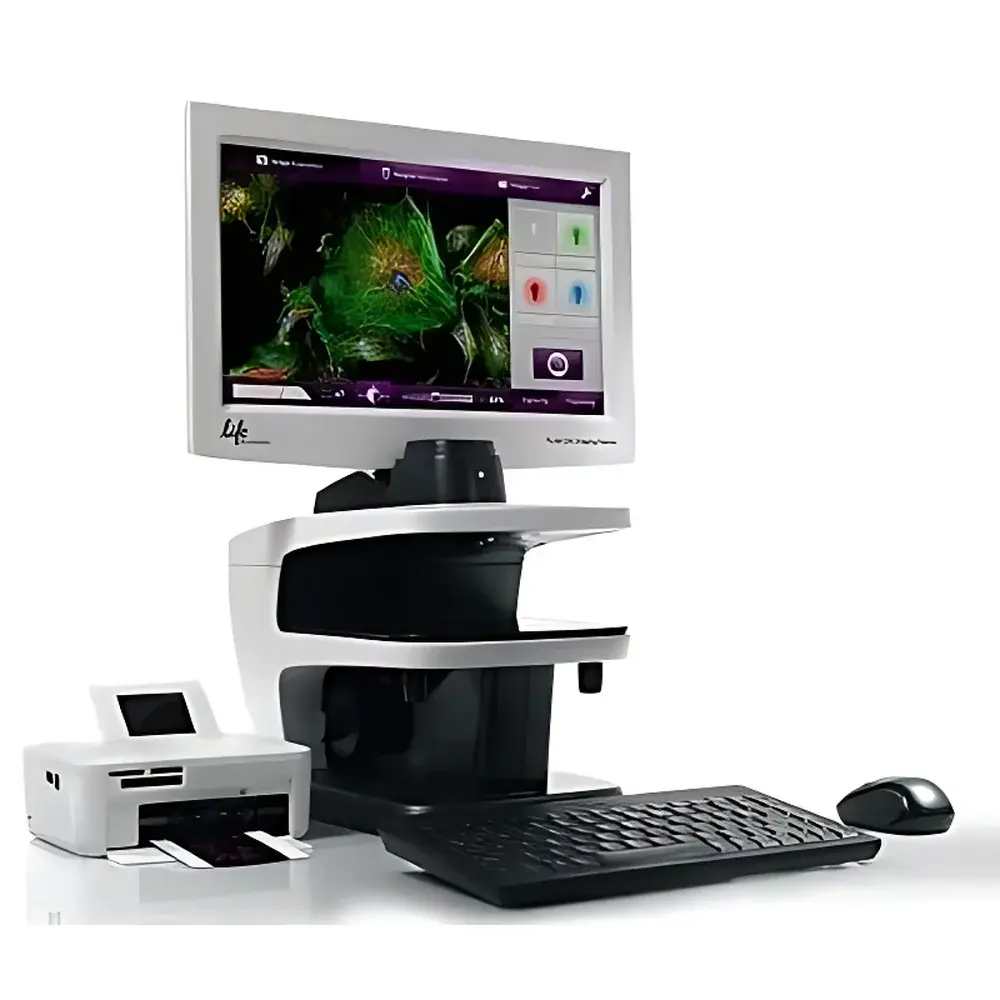
| Display | 15″ color LCD, 1366 × 768 resolution, adjustable tilt |
| Objective | 20× Plan Fluor, NA 0.45, WD 5.9 mm |
| Total Magnification | 460× (optical) to 1840× (digital zoom) |
| Resolution Limit | 0.5 µm |
| Illumination | Long-life LED (50,000 h), intensity-adjustable |
| Excitation Filters | 390/40 nm (blue), 482/18 nm (green), 586/15 nm (red) |
| Emission Filters | 446/33 nm (blue), 532/59 nm (green), 646/68 nm (red) |
| Imaging Modes | Brightfield (relief contrast phase), Blue/Green/Red fluorescence |
| Channels | 4 (phase + 3 fluorescence) |
| Stage Travel | X/Y ±4 mm |
| Stage Clearance | 60 mm |
| Supported Vessels | Slides, multiwell plates, culture flasks |
| File Formats | 16-bit monochrome TIFF, JPEG, BMP, PNG |
| Software | FLoid™ Cell Imaging Station Software (v3.x or later) |
| Language Support | English, Spanish, German, French, Italian, Japanese, Chinese |
| USB Ports | 4 |
| Included Media | 2 GB USB drive |
Overview
The Invitrogen EVOS FLoid™ Cell Imaging Station is a benchtop, integrated fluorescence and brightfield imaging system engineered for rapid, reproducible acquisition of high-fidelity cellular images in standard laboratory environments—without requiring darkroom operation or complex optical alignment. Built on a fixed-optics platform with precisely aligned LED excitation sources and bandpass-filtered detection pathways, the FLoid system employs widefield epifluorescence microscopy principles to deliver consistent signal-to-noise performance across all four imaging channels: relief contrast phase, blue (DAPI/Hoechst), green (FITC/GFP), and red (TRITC/mCherry). Its compact, self-contained architecture eliminates the need for external light sources, filter wheels, or camera controllers—reducing variability and simplifying workflow validation. Designed for routine cell culture monitoring, transfection assessment, and viability screening, the FLoid station operates as a dedicated, GxP-adjacent imaging tool that complements—but does not replace—high-resolution confocal or super-resolution platforms.
Key Features
- Intuitive, single-touch interface: Proprietary FLoid software features a streamlined, icon-driven workflow enabling image capture, channel blending, annotation, and export in under 90 seconds—no prior microscopy training required.
- Benchtop fluorescence without darkroom dependency: Optimized LED illumination with thermally stabilized output and precision dichroic beam splitting ensures stable excitation intensity and minimal photobleaching during repeated acquisitions.
- Modular stage design: 60 mm vertical clearance and ±4 mm XY travel accommodate diverse sample formats—including standard glass slides, 6–384-well plates, T25/T75 flasks, and Petri dishes—enabling direct imaging of adherent and suspension cultures.
- Calibrated optical path: Fixed 20× Plan Fluor objective (NA 0.45, WD 5.9 mm) provides uniform field illumination and diffraction-limited lateral resolution of ≤0.5 µm at 550 nm, validated per ISO 19012-1 for objective performance.
- Regulatory-ready data handling: All acquired images are saved in 16-bit TIFF format with embedded metadata (timestamp, objective ID, exposure time, gain, channel configuration), supporting traceability requirements under GLP and internal QA protocols.
Sample Compatibility & Compliance
The FLoid system supports a broad range of biological specimens—from fixed and live mammalian cells to yeast, bacteria, and tissue sections—when used with compatible Molecular Probes® reagents (over 160 validated dyes and probes documented in Thermo Fisher’s online assay database). Its open-stage architecture permits imaging through standard tissue-culture-treated plastic and glass-bottom vessels without immersion media or coverslip correction. While not certified for clinical diagnostics (IVD), the instrument meets IEC 61010-1 safety standards for laboratory equipment and complies with FCC Part 15 Class B and CE electromagnetic compatibility directives. Its firmware and software architecture support audit trail generation and user-access controls—facilitating alignment with FDA 21 CFR Part 11 expectations for electronic records when deployed within controlled environments.
Software & Data Management
The FLoid™ Cell Imaging Station Software (v3.x or later) runs natively on Windows-based host systems and includes built-in tools for real-time channel overlay, histogram-based exposure optimization, region-of-interest (ROI) intensity quantification, and batch export to CSV or PDF. Image metadata—including objective magnification, LED intensity percentage, exposure duration (1–5000 ms), and gain settings—is automatically appended to each TIFF header. The included 2 GB USB drive contains software installers, quick-start guides, and reference protocols; users may configure network storage destinations via SMB/CIFS mounts. Language localization (English, Spanish, German, French, Italian, Japanese, Chinese) ensures operational consistency across multinational research sites. No cloud connectivity or telemetry is enabled by default—data residency remains fully under institutional control.
Applications
- Quantitative assessment of transfection efficiency using fluorescent protein reporters (e.g., GFP, RFP)
- Routine confluence and morphology monitoring in stem cell and primary culture workflows
- Cell viability assays (e.g., Calcein AM/EthD-1, Hoechst/PI) with dual-channel ratiometric analysis
- Immunofluorescence screening of fixed samples labeled with Alexa Fluor® or DyLight® conjugates
- Time-lapse imaging of dynamic processes (e.g., wound healing, mitosis) with programmable interval capture
- Documentation and archiving of QC checkpoints in bioproduction and cell banking operations
FAQ
Is the FLoid system compatible with live-cell imaging over extended durations?
Yes—its low-heat LED illumination and absence of mercury/xenon lamp cycling minimize thermal drift and phototoxicity. However, environmental control (CO₂, temperature, humidity) must be provided externally via incubation chambers or stage-top systems.
Can third-party fluorophores be used with the FLoid’s filter sets?
The excitation/emission bands are optimized for common dyes (DAPI, FITC, TRITC); spectral overlap should be verified using Thermo Fisher’s online fluorophore spectra tool before adoption of novel probes.
Does the system support Z-stack acquisition or autofocus?
No—the FLoid is a fixed-focus, single-plane imaging platform. For axial sectioning or focus stabilization, users should integrate it with compatible motorized stages or external autofocus add-ons.
What regulatory documentation is available for installation qualification (IQ) and operational qualification (OQ)?
Thermo Fisher provides a Factory Acceptance Test (FAT) report and optional IQ/OQ protocol templates aligned with ASTM E2500 and ISO/IEC 17025 guidelines—available upon request through certified service partners.
How is software updated and validated after deployment?
Updates are distributed via secure download; each release includes version-controlled release notes and verification test reports. Institutions may perform local validation using predefined image quality metrics (e.g., MTF, SNR, uniformity) per internal SOPs.


