IPX-LC Integrated Imaging Plate-Based X-ray Laue Camera
| Origin | Japan |
|---|---|
| Manufacturer Type | Distributor |
| Origin Category | Imported |
| Model | IPX-LC |
| Pricing | Upon Request |
Overview
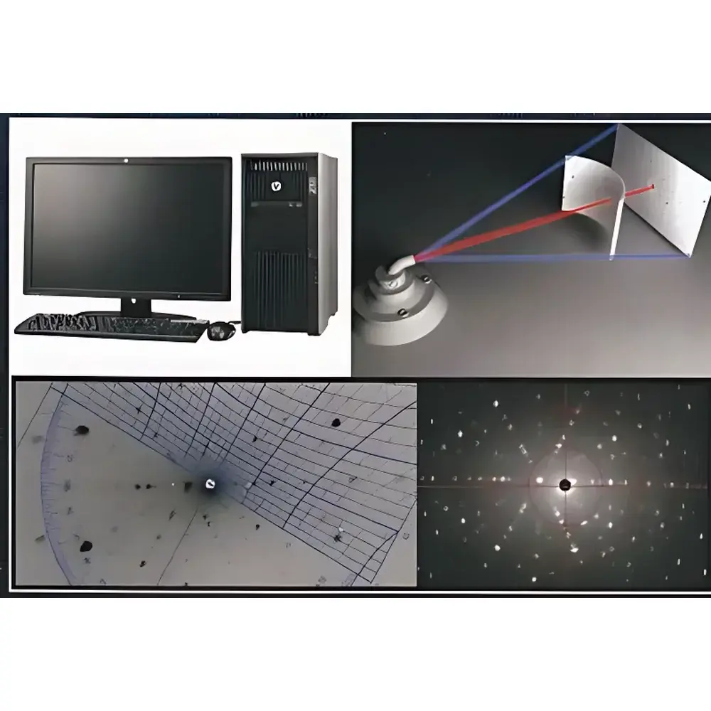
The IPX-LC Integrated Imaging Plate-Based X-ray Laue Camera is a precision single-crystal orientation analysis system engineered for laboratory-based Laue diffraction measurements. It operates on the fundamental principle of Laue diffraction—using polychromatic (white) X-ray radiation incident on a stationary single crystal, where Bragg’s law (nλ = 2d sinθ) governs wavelength-selective reflection from crystallographic planes (hkl). Unlike monochromatic techniques such as rotating anode or synchrotron-based methods, the Laue method captures simultaneous diffraction from multiple lattice planes in a single exposure, enabling rapid determination of crystal orientation, misorientation angles, and incident beam geometry relative to the crystal frame. Designed for high reproducibility and operational simplicity, the IPX-LC replaces conventional photographic film with reusable phosphor-based imaging plates (IP), integrating exposure, laser scanning readout, and optical erasure into a single automated workflow. Its compact, benchtop architecture supports integration with standard 2 kW sealed-tube X-ray generators equipped with tungsten (W) anodes, making it suitable for routine quality control and R&D environments in semiconductor manufacturing, quartz resonator calibration, and advanced aerospace material characterization.
Key Features
- Integrated imaging plate (IP) detection system with fully automated exposure–readout–erase cycle, eliminating consumable film and darkroom processing
- Cylindrical 120° IP holder accommodating 120 mm × 80 mm phosphor plates, optimized for uniform angular coverage in back-reflection Laue geometry
- High-resolution laser scanner with selectable pixel sizes: 100 µm (standard), 200 µm (fast mode), and 50 µm (high-resolution mode over 50 mm × 50 mm subregion)
- Motorized dual-axis goniometer (α, β ±20°; rotation axis 360°) enabling precise crystal alignment without repositioning the main camera body
- Interchangeable collimators (Φ0.5 mm and Φ1.0 mm) mounted on fixed ports—no mechanical realignment required during switching
- Fixed camera center height of 300 mm above the stage for repeatable setup across multiple samples and users
- Real-time image preview and post-acquisition processing supported by dedicated Windows-based software suite
Sample Compatibility & Compliance
The IPX-LC accommodates a wide range of single-crystalline materials including silicon, sapphire, quartz, gallium arsenide (GaAs), lithium niobate (LiNbO₃), and nickel-based superalloys. Its design conforms to ISO 17025-relevant practices for measurement traceability in materials testing laboratories. While not certified as medical or industrial radiography equipment under IEC 61331-1, the system complies with general radiation safety guidelines when operated with approved X-ray generators and shielding enclosures. All software modules support audit-trail logging and user-access controls compatible with GLP/GMP documentation requirements. Data export formats (TIFF, ASCII, CIF-compatible orientation matrices) facilitate interoperability with third-party crystallographic analysis tools such as MTEX, OIM Analysis, and JADE.
Software & Data Management
The IPX-LC is delivered with three core software applications running natively on Windows XP or Vista platforms: IP-Xray for real-time IP readout and intensity mapping; TRY-Viewer for interactive LAUE spot annotation, background subtraction, and geometric calibration; and TRY-Converter, which performs cylindrical-to-Cartesian coordinate transformation essential for accurate pole figure reconstruction and Euler angle calculation. All software modules store metadata—including exposure time, collimator ID, goniometer angles, and IP batch number—in embedded file headers. Raw image data are stored losslessly in 16-bit TIFF format, supporting batch processing and scripting via COM interface for integration into automated QA/QC pipelines.
Applications
- Determination of crystallographic orientation and misorientation in semiconductor wafers and epitaxial layers
- Calibration of quartz crystal resonators used in frequency control devices
- Orientation mapping of turbine blade single-crystal superalloys for creep resistance validation
- Residual stress estimation via Laue peak splitting analysis (when combined with calibrated strain standards)
- Educational demonstration of reciprocal lattice geometry and Bragg condition visualization
- Pre-screening tool prior to high-resolution synchrotron or neutron diffraction experiments
FAQ
What type of X-ray source is compatible with the IPX-LC?
The system is designed for use with standard laboratory 2 kW sealed-tube X-ray generators equipped with tungsten (W) anodes and appropriate high-voltage power supplies (typically 20–60 kV). Synchrotron or microfocus sources are not supported due to beam geometry and intensity constraints.
Can the IP plates be reused indefinitely?
Yes—phosphor imaging plates are rated for >10,000 read/erase cycles when handled according to manufacturer specifications and stored in low-humidity, light-shielded conditions.
Is the IPX-LC compliant with FDA 21 CFR Part 11?
While the software includes user authentication and basic audit trail functionality, full Part 11 compliance requires additional validation protocols and electronic signature implementation, which must be performed by the end-user laboratory per internal SOPs.
Does the system support automatic indexing of Laue patterns?
No—the IPX-LC provides high-fidelity raw diffraction images and geometric transformation tools; indexing and orientation solution require manual or semi-automated input using external crystallographic databases or third-party indexing algorithms.
What is the maximum usable exposure time for the IP plates?
Exposure duration depends on X-ray flux and sample absorption; typical ranges are 1–300 seconds. Overexposure may cause saturation or nonlinearity—users are advised to perform preliminary test exposures using attenuated beams.