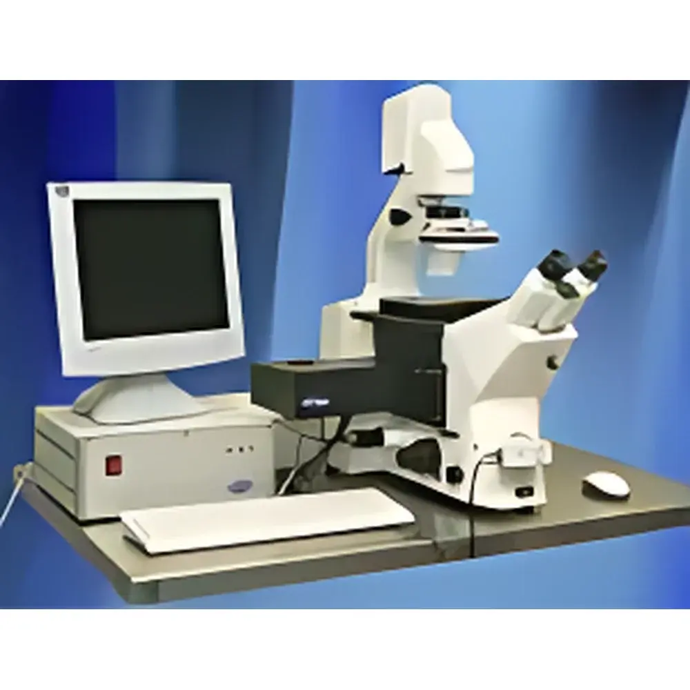
JenLab TauMap® Fluorescence Lifetime Imaging Microscope
| Brand | JenLab |
|---|---|
| Origin | Germany |
| Model | TauMap® |
| Type | FLIM/FCS/FRET Multimodal Microscopy System |
| Application Domain | Biomedical Research, Pharmaceutical Development, Live-Cell Dynamics |
Overview
The JenLab TauMap® Fluorescence Lifetime Imaging Microscope is a turnkey multimodal platform engineered for quantitative, time-resolved fluorescence analysis at the single-cell and subcellular level. Based on time-correlated single-photon counting (TCSPC) and pulsed near-infrared laser excitation, the system delivers picosecond-resolution fluorescence lifetime measurements across multiple spectral channels. Unlike intensity-based fluorescence microscopy, FLIM provides intrinsic contrast independent of fluorophore concentration, photobleaching, or excitation intensity—making it ideal for quantitative mapping of molecular microenvironments, metabolic states (e.g., NAD(P)H/FAD redox ratios), and conformational dynamics in living specimens. Integrated FCS capability enables diffusion coefficient quantification and molecular brightness analysis, while FRET efficiency mapping supports real-time interrogation of protein–protein interactions, ligand binding, and conformational changes with nanometer-scale spatial sensitivity.
Key Features
- TCSPC-based FLIM acquisition with 10⁶ photon/s detection throughput
- Integrated femtosecond fiber laser (780–860 nm tunable, 80 MHz repetition rate) with dispersion compensation for deep-tissue two-photon excitation
- Simultaneous dual-channel spectral detection (e.g., 400–500 nm and 500–650 nm) with high quantum-efficiency hybrid PMTs
- Motorized XYZ stage with piezo-driven Z-focus for volumetric lifetime tomography up to 500 µm depth in scattering tissue
- Real-time FLIM data reconstruction using GPU-accelerated phasor analysis and multi-exponential decay fitting (mono-, bi-, and tri-exponential models)
- FCS module with autocorrelation analysis (τ = 10 µs–1 s) and cross-correlation for co-diffusion studies
- FRET quantification via donor lifetime quenching analysis—no acceptor photobleaching required
Sample Compatibility & Compliance
The TauMap® accommodates live biological specimens ranging from adherent cultured cells and 3D organoids to ex vivo tissue slices and intravital preparations in murine models. Its non-descanned detection path and low-photon-dose imaging protocol minimize phototoxicity during longitudinal experiments. The system complies with ISO 13485 design control principles for research-grade instrumentation and supports audit-ready documentation workflows aligned with GLP and GMP preclinical study requirements. All software modules adhere to FDA 21 CFR Part 11 electronic record and signature standards, including user access controls, electronic audit trails, and immutable data archiving. Instrument calibration is traceable to NIST-certified reference dyes (e.g., Rhodamine B, Fluorescein) and validated per ASTM E2879-21 for FLIM system performance verification.
Software & Data Management
TauMap® is operated via JenLab’s proprietary TauFit software suite—a modular, scriptable environment built on Qt/C++ with Python API integration. Core capabilities include automated lifetime histogram generation, pixel-wise decay model selection, phasor plot navigation with region-of-interest (ROI) gating, and batch processing of time-lapse FLIM stacks. FCS data undergoes rigorous noise filtering, triplet-state correction, and diffusion model fitting with confidence interval estimation. Export formats include HDF5 (with metadata schema compliant with OME-NGFF), TIFF (32-bit float lifetime maps), and CSV (for statistical analysis in R or MATLAB). Data integrity is enforced through SHA-256 checksums, versioned project files, and optional integration with institutional LIMS or ELN systems via RESTful API.
Applications
- Mitochondrial metabolism profiling: Quantitative mapping of free/bound NADH lifetime components to assess oxidative phosphorylation status in cancer spheroids and iPSC-derived cardiomyocytes
- Protein interaction kinetics: Time-resolved FRET between GFP-tagged receptors and mCherry-labeled effectors during GPCR internalization in primary neurons
- Drug-target engagement: In situ measurement of target residence time via fluorescent probe lifetime shifts in live tumor xenografts
- Membrane microdomain dynamics: FCS-based diffusion coefficient gradients across lipid raft boundaries in stimulated T-cells
- Neurovascular coupling: Simultaneous two-photon FLIM of Ca²⁺ indicators and hemoglobin oxygenation in cortical capillaries during functional stimulation
FAQ
What laser wavelengths are supported for two-photon excitation?
The integrated fiber laser operates tunably from 780 nm to 860 nm; optional OPO extension enables coverage up to 1300 nm for deeper penetration.
Can TauMap® be integrated with existing confocal or multiphoton microscope frames?
Yes—JenLab offers OEM-compatible TCSPC detection modules and synchronization interfaces (TTL, LVDS) for retrofitting into Zeiss LSM, Leica SP, or Bruker Ultima platforms.
Is lifetime calibration traceable to international standards?
Yes—calibration uses NIST-traceable reference dyes and follows procedures defined in ISO/IEC 17025-accredited laboratories; full calibration reports are provided with each system shipment.
Does the system support real-time FLIM during animal surgery?
Yes—low-latency acquisition (<50 ms/frame at 256×256) and embedded phasor analysis enable intraoperative metabolic imaging with immediate visual feedback.
How is photodamage minimized during long-term live-cell imaging?
By combining adaptive illumination power control, gated detection windows, and high-sensitivity detectors, average irradiance remains below 5 mW/mm²—well within established phototoxicity thresholds for mammalian cells (per ISO 21542:2021).


