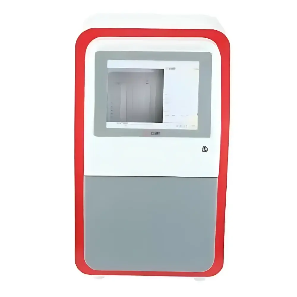
Jiapeng JP-K3300Plus Chemiluminescence Gel Imaging System
| Brand | Jiapeng |
|---|---|
| Origin | Shanghai, China |
| Model | JP-K3300Plus |
| Instrument Type | Chemiluminescence Gel Imaging System |
| CCD Resolution | 9 MP (3000 × 3000) |
| Bit Depth | 16-bit |
| Dynamic Range | 4.8 OD (0–65535 grayscale) |
| Pixel Size | 5.4 µm × 5.4 µm |
| Sensor Format | 4/3-inch Sony IMX533CLK-D |
| Cooling Temperature | −45 °C (ΔT ≤ −65 °C vs ambient) |
| Lens Aperture | f/0.95 fixed-focus lens |
| Signal-to-Noise Ratio | ≥56 dB |
| Quantum Efficiency | >75% |
| White Light Source | Top-mounted uniform LED transilluminator |
| Dual Imaging Area | 9.5 × 9.5 cm & 12.5 × 12.5 cm |
| Power Supply | AC 220 V, 50/60 Hz |
| Dimensions (W×D×H) | 390 × 440 × 735 mm |
| Weight | 18.3 kg |
Overview
The Jiapeng JP-K3300Plus Chemiluminescence Gel Imaging System is a fully automated, high-sensitivity digital imaging platform engineered for quantitative detection and analysis of nucleic acids and proteins in gels and membranes. It operates on the principle of low-light photon capture using a thermoelectrically cooled scientific CMOS sensor, optimized for chemiluminescent, bioluminescent, and fluorescent signals—particularly those generated by horseradish peroxidase (HRP) or alkaline phosphatase (AP) substrate reactions. Unlike conventional UV transilluminators or basic CCD-based systems, the JP-K3300Plus integrates hardware-level cooling, ultra-fast optics, and multi-area sample positioning to deliver reproducible, background-suppressed imaging across diverse assay formats—including Western blots, Southern/Northern blots, EMSA, and DNA ladders stained with ethidium bromide or SYBR dyes.
Key Features
- Scientific-grade 4/3-inch Sony IMX533CLK-D sensor with active cooling to −45 °C (up to 65 °C below ambient), minimizing dark current (<0.0005 e⁻/pixel/sec) and enabling long-exposure acquisition without thermal noise accumulation.
- f/0.95 fixed-focus lens with high transmission efficiency and minimal chromatic aberration—optimized for broad-spectrum emission from luminol-based substrates and near-infrared fluorophores.
- Dual-position drawer-style sample stage supporting two distinct imaging areas (9.5 × 9.5 cm and 12.5 × 12.5 cm), facilitating side-by-side comparison of multiple membranes or sequential imaging under varying exposure conditions.
- Top-mounted uniform white LED transilluminator with calibrated intensity control, designed to meet ISO 15775 requirements for gel documentation consistency and enabling accurate normalization against loading controls.
- User-configurable auto-shutdown timer (0–60 min) for all light sources, reducing photobleaching risk and extending reagent shelf life during extended protocol runs.
- Integrated 12-inch industrial-grade touchscreen PC with preloaded imaging software—eliminating external computer dependency and ensuring deterministic USB 3.0 data transfer latency.
Sample Compatibility & Compliance
The JP-K3300Plus accommodates standard electrophoretic formats including mini- and midi-gels (up to 15 × 15 cm), nitrocellulose and PVDF membranes (up to 13 × 13 cm), and microplates (96-well). Its optical design conforms to IEC 61000-6-3 EMC standards and includes built-in light-tight shielding compliant with ISO 17025 laboratory environmental control guidelines. While not FDA 21 CFR Part 11-certified out-of-the-box, the system supports audit-trail-enabled software upgrades compatible with GLP/GMP workflows when deployed with validated SOPs and instrument qualification protocols (IQ/OQ/PQ).
Software & Data Management
Bundled imaging software provides real-time preview, multi-channel overlay (chemiluminescence + white light reference), background subtraction via rolling-ball algorithm, and lane/band quantification using integrated density profiling. Export options include TIFF (uncompressed), PNG, and CSV-formatted intensity matrices suitable for downstream statistical analysis in GraphPad Prism or R. All image metadata—including exposure time, lens aperture, sensor temperature, and gain settings—is embedded in EXIF tags, satisfying traceability requirements under ISO/IEC 17025 Clause 7.5.2.
Applications
- Quantitative Western blotting with HRP-conjugated secondary antibodies and enhanced chemiluminescent substrates (e.g., Luminata Forte, SuperSignal West Femto).
- DNA/RNA gel documentation with sub-nanogram sensitivity (≤0.01 ng EB-stained DNA detectable at 1×1 binning mode).
- Reporter gene assays involving luciferase-based bioluminescence in cell lysates or live-cell imaging plates.
- Protein interaction studies via chemiluminescent proximity ligation assays (PLA) and in-gel zymography.
- Quality control of CRISPR-Cas9 editing efficiency through cleavage product visualization on high-resolution polyacrylamide gels.
FAQ
What is the minimum detectable signal level for HRP-mediated chemiluminescence?
The system achieves reliable detection of ≤1 pg of horseradish peroxidase under optimized substrate conditions using 5-minute exposures at −45 °C sensor temperature and f/0.95 lens.
Can the JP-K3300Plus be used for fluorescence imaging?
Yes—when equipped with optional excitation/emission filter sets (e.g., FITC, TRITC, Cy5), it supports multiplex fluorescent blotting; however, dedicated fluorescence performance requires validation against NIST-traceable standards.
Is remote operation supported?
The onboard industrial PC supports VNC and RDP protocols; secure remote access may be configured within institutional firewall policies, though raw image streaming is limited to local network segments for bandwidth preservation.
How is calibration maintained across instruments?
Each unit ships with a certified neutral-density reference slide traceable to NIM (National Institute of Metrology, China); annual recalibration services are available through authorized Jiapeng service centers.
Does the software support batch processing of multiple blots?
Yes—the workflow manager enables scripted acquisition sequences, automatic file naming based on user-defined templates, and parallel quantification across up to 16 lanes per image with inter-blot normalization anchors.


