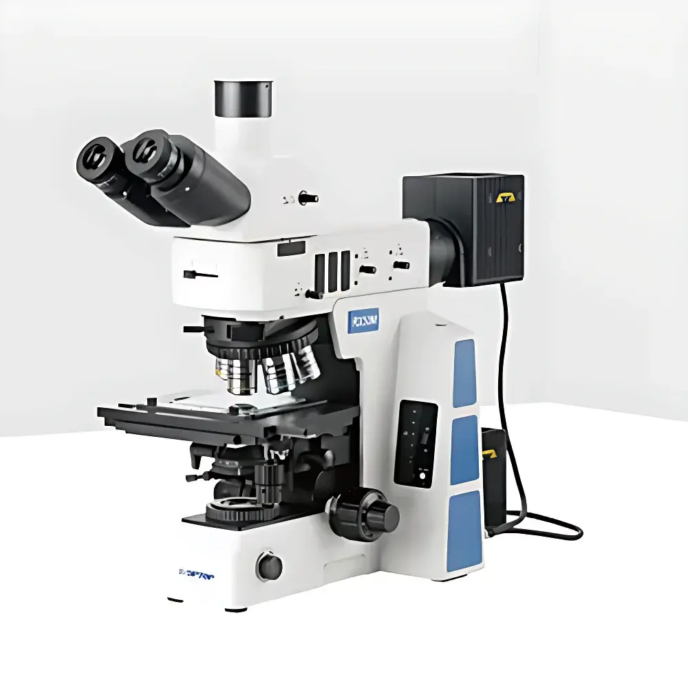
Jihepu RX50M Research-Grade Inverted Metallurgical Microscope
| Brand | Jihepu |
|---|---|
| Origin | Shandong, China |
| Model | RX50M |
| Type | Inverted Metallurgical Microscope |
| Total Magnification | 26.5× (with 10× eyepiece and 2.65× auxiliary lens, or standard optical magnification up to 1000× with 100× objective) |
| Eyepieces | Wide-field Plan Eyepieces PL10×/25 mm and PL10×/26.5 mm, adjustable diopter, optional crosshair reticle |
| Objectives | Semi-Apochromatic Brightfield/Darkfield Metallurgical Objectives (5×, 10×, 20×, 50×, 100× oil) |
| Nosepiece | 5-position or 6-position Brightfield/Darkfield Turret with DIC slot |
| Illumination | 12 V / 100 W Halogen Lamp, Digital Intensity Control, Dual Path (Reflected & Transmitted), Built-in Filter Slots (LBD, ND6, ND25), Polarizer/Analyzer Mounts |
| Stage | 4-inch Mechanical Stage (105 × 102 mm travel), Y-axis lock, optional transmission shutter and glass plate |
| Focus Mechanism | Coaxial Coarse/Fine Focus, 25 mm coarse travel, 1 µm fine step resolution, anti-slip tension adjustment and upper limit stop |
| Optical System | Infinity-Corrected, Achromatic Condenser (NA 0.9), Optional DIC and Interference Filters (Blue ≤480 nm, Green 520–570 nm, Red 630–750 nm, White Balance Plate) |
| Image Analysis System | Integrated Digital Imaging Interface (C-mount, 0.5× reduction lens), Software-ready for quantitative metallographic analysis |
| Compliance | Designed to support ISO 4287, ASTM E3, ASTM E112, ISO 643, and EN 10360-2 workflows |
Overview
The Jihepu RX50M is a research-grade inverted metallurgical microscope engineered for high-precision microstructural characterization of metallic, ceramic, and composite specimens in industrial quality control, failure analysis, and academic metallurgy laboratories. Its inverted configuration positions the objective lenses beneath the specimen stage—ideal for observing large, heavy, or irregularly shaped samples such as castings, weldments, or mounted cross-sections without reorientation. The system employs an infinity-corrected optical path with semi-apochromatic objectives optimized for both brightfield and darkfield illumination, minimizing chromatic and spherical aberrations across the full magnification range (5× to 1000×). Coupled with a 26.5 mm field number wide-field eyepiece system and a high-transmission halogen illumination source, the RX50M delivers exceptional image flatness, contrast uniformity, and color fidelity—critical for grain size measurement, phase identification, inclusion rating, and porosity analysis per ASTM E112 and ISO 643 standards.
Key Features
- Inverted optical architecture with low-handled coaxial focus controls, enabling ergonomic operation during extended observation sessions.
- Three-way trinocular head with selectable beam-splitting ratios (100:0, 20:80, or 0:100) for simultaneous visual inspection and digital documentation—supporting real-time side-by-side comparison of live and recorded imagery.
- Five- or six-position objective turret compatible with differential interference contrast (DIC) modules and equipped with dedicated slots for polarizers and analyzers—enabling quantitative birefringence assessment and stress-induced anisotropy mapping.
- Digital-intensity-controlled 12 V / 100 W halogen illumination with memory-based brightness presets, reflected/transmitted light toggle, and integrated neutral density (ND6, ND25) and LBD filters for optimal contrast tuning across diverse sample reflectivities.
- Mechanical 4-inch stage with 105 mm × 102 mm travel range, Y-axis locking mechanism, and optional transmission-mode shutter—facilitating repeatable positioning and multi-site comparative analysis on serial sections.
- Substage achromatic condenser (NA 0.9) with centerable aperture and field diaphragms, plus interchangeable interference filters (blue, green, red, white balance) for spectral optimization in etched or unetched surface evaluation.
Sample Compatibility & Compliance
The RX50M accommodates standard metallographic specimens up to 30 mm in height and 150 mm in diameter when using optional stage extensions. Its robust inverted frame supports polished, coated, or bulk specimens—including those requiring vacuum-compatible mounting or thermal stabilization stages (externally interfaced). The system complies with core metrological requirements outlined in ISO/IEC 17025 for calibration traceability and aligns with analytical workflows defined in ASTM E3 (preparation of metallographic specimens), ASTM E112 (grain size determination), and ISO 4287 (surface roughness profiling via optical sectioning). Optional DIC and polarization accessories extend conformance to ISO 10816-3 (vibration-induced microcrack detection) and EN 10360-2 (dimensional metrology of microstructures).
Software & Data Management
The RX50M integrates seamlessly with third-party image analysis platforms via C-mount interface and 0.5× reduction optics, ensuring pixel-level registration between optical magnification and sensor resolution. Its hardware design supports time-stamped image capture, Z-stack acquisition, and region-of-interest (ROI) annotation—features essential for GLP-compliant reporting and FDA 21 CFR Part 11–aligned audit trails when paired with validated software suites. The microscope’s digital illumination controller enables programmable light intensity profiles synchronized with automated stage movement—critical for batch-mode analysis of production lots under consistent photometric conditions.
Applications
- Quantitative metallography: grain boundary delineation, second-phase particle counting, and carbide distribution mapping in steels and superalloys.
- Weld integrity assessment: heat-affected zone (HAZ) microstructure evaluation, fusion line detection, and porosity quantification per AWS D1.1.
- Failure analysis: fracture surface topography correlation with crack propagation direction, inclusion morphology classification (ASTM E45), and decarburization depth measurement.
- Coating and plating inspection: thickness uniformity verification, interfacial delamination detection, and diffusion layer characterization in thermal spray or electroplated components.
- Research in additive manufacturing: powder morphology screening, melt pool geometry analysis, and residual stress visualization using polarized light and DIC contrast enhancement.
FAQ
Is the RX50M compatible with motorized stage and autofocus systems?
Yes—the trinocular port and standardized mechanical interfaces support OEM integration of motorized XY stages and hardware-triggered autofocus modules for automated tile scanning and focus stacking.
Does the system meet regulatory requirements for pharmaceutical or medical device QC labs?
While primarily designed for metallurgical applications, its optical stability, illumination reproducibility, and digital output architecture enable validation under ISO 13485 and GMP Annex 11 when deployed with qualified software and documented IQ/OQ protocols.
Can darkfield imaging be performed on non-conductive or coated samples?
Yes—the dedicated darkfield reflector and adjustable aperture diaphragm allow high-contrast edge enhancement on anodized aluminum, PVD-coated tools, or graphite-embedded composites without conductive coating.
What is the maximum working distance achievable with the 100× objective?
The 100× oil-immersion semi-apochromatic objective provides a working distance of 0.12 mm; dry alternatives (e.g., 50× long-working-distance objective) offer 5.1 mm WD for thick-section or encapsulated sample observation.
Are calibration certificates available for the included micrometers and stage encoders?
Factory-calibrated stage encoders and high-accuracy stage micrometers (0.01 mm division) are supplied with NIST-traceable calibration reports upon request—required for ISO/IEC 17025 accreditation of measurement uncertainty budgets.


