KJ GROUP BT-1600 Image-Based Particle Analysis System
| Brand | KJ GROUP |
|---|---|
| Origin | Liaoning, China |
| Manufacturer Type | Authorized Distributor |
| Country of Origin | China |
| Model | BT-1600 |
| Price | Upon Request |
| Measurement Range | 1–3000 µm |
| Repeatability Error | ≤3% |
| Maximum Magnification | ≤4000× |
| Optical Resolution | ≤0.1 µm |
| CCD Camera | 5 MP |
| Microscope Options | Domestic, Nikon, or Metallographic |
| Calibration Scale | 10 µm |
| Analyzable Parameters | Particle Size Distribution, Aspect Ratio (Length/Width), Circularity Distribution, Individual & Ensemble Particle Morphology |
| Automated Segmentation Speed | <1 s per image |
| Segmentation Success Rate | >93% |
Overview
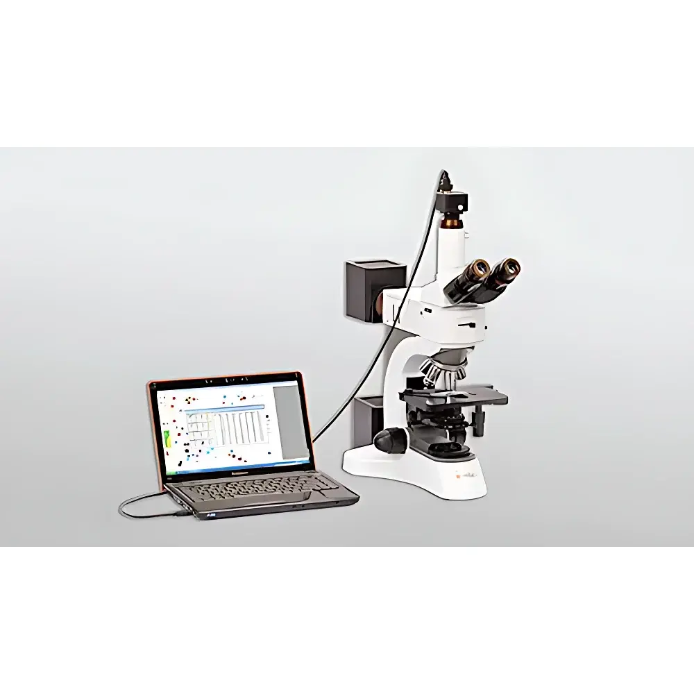
The KJ GROUP BT-1600 Image-Based Particle Analysis System is an integrated optical-morphological characterization platform engineered for quantitative microstructural analysis of dispersed particulate systems. It operates on the principle of high-resolution digital microscopy coupled with algorithm-driven binary segmentation and geometric feature extraction. Unlike ensemble-averaging techniques such as laser diffraction or dynamic light scattering, the BT-1600 delivers true two-dimensional morphometric data—including equivalent circular diameter, Feret diameters, aspect ratio, convexity, and circularity—for each resolved particle within a field of view. This enables statistically robust population-level inference while preserving individual particle identity—a critical requirement in quality control of anisotropic, irregular, or aggregated materials where shape directly influences functional performance (e.g., flowability, packing density, reactivity, or optical scattering). The system is calibrated traceably to NIST-traceable stage micrometers and conforms to foundational metrological practices outlined in ISO 13322-1:2014 (Particle size analysis — Image analysis methods — Part 1: Static image analysis methods).
Key Features
- Modular optical architecture supporting interchangeable microscope configurations—standard upright, inverted, or metallographic variants—with optional Nikon objectives (up to 100× oil immersion) for sub-micron resolution imaging.
- Dedicated 5-megapixel progressive-scan CCD camera with 12-bit dynamic range, USB 3.0 interface, and hardware-triggered acquisition to minimize motion blur during rapid sample scanning.
- Automated calibration workflow incorporating real-time scale validation via embedded 10 µm reference graticule; supports multi-point spatial correction across the full field of view.
- Real-time segmentation engine employing adaptive thresholding, morphological noise suppression, and watershed-based splitting of touching particles—achieving >93% success rate even for dense, overlapping clusters typical in milled ceramics or agglomerated battery cathodes.
- Batch processing capability for unattended analysis of multi-field image stacks, with configurable pass/fail criteria based on user-defined morphological thresholds (e.g., aspect ratio >5 for needle-like silicates).
- Rugged mechanical design with vibration-damped base plate and precision XYZ motorized stage (optional), enabling reproducible positioning for time-series or comparative studies under GLP-compliant workflows.
Sample Compatibility & Compliance
The BT-1600 accommodates dry powders, suspended slurries (aqueous or organic), and fixed biological specimens mounted on standard 1″ × 3″ glass slides. Sample preparation follows ASTM D7216-18 guidelines for dry dispersion and ISO 13322-2:2020 protocols for liquid suspension stability assessment. The system supports compliance documentation generation—including audit trails, electronic signatures, and version-controlled analysis templates—aligned with FDA 21 CFR Part 11 requirements when deployed in regulated pharmaceutical or medical device manufacturing environments. All measurement algorithms are validated against certified reference materials (e.g., NIST SRM 1980, monodisperse latex spheres) and documented per ISO/IEC 17025:2017 analytical method validation clauses.
Software & Data Management
The proprietary BT-Analysis Suite v4.x provides a deterministic, scriptable environment for morphometric quantification. It includes built-in modules for statistical outlier detection (Grubbs’ test), cumulative distribution fitting (Rosin-Rammler, log-normal), and multivariate correlation mapping (e.g., circularity vs. area). Raw images, processed masks, and metadata (timestamp, operator ID, instrument settings) are stored in vendor-neutral TIFF + XML sidecar format. Export options include CSV (for Excel/Python integration), PDF reports with embedded histograms and scatter plots, and direct SQL database push to LIMS platforms via ODBC drivers. Software validation packages—including IQ/OQ documentation and change control logs—are available upon request for GMP-regulated deployments.
Applications
- Abrasive manufacturing: Quantifying angularity and fracture surface morphology of SiC, Al₂O₃, and diamond grits to predict cutting efficiency and tool wear.
- Lithium-ion battery R&D: Measuring sphericity and surface roughness of spherical graphite anode powders—parameters directly correlated with electrode slurry rheology and first-cycle Coulombic efficiency.
- Advanced metallurgy: Characterizing dendritic arm spacing and secondary phase distribution in atomized aluminum alloys for additive manufacturing feedstock qualification.
- Pharmaceutical excipients: Assessing particle elongation and surface texture of microcrystalline cellulose grades to model compaction behavior during tablet formulation.
- Geological and environmental science: Differentiating biogenic vs. lithogenic silt fractions in sediment cores using convexity and edge complexity metrics.
- Food science: Monitoring starch granule swelling kinetics and gelatinization-induced morphological transitions in real time via time-lapse imaging mode.
FAQ
What magnification is required to resolve particles at the lower end of the 1–3000 µm range?
At 1 µm resolution, a minimum optical magnification of 100× (with 10× eyepiece and 10× objective) is recommended, paired with appropriate numerical aperture (NA ≥ 0.25) and Köhler illumination alignment.
Can the system analyze transparent or low-contrast particles such as polymer beads?
Yes—via optional differential interference contrast (DIC) or darkfield illumination modules; software includes phase-enhancement filters optimized for low-contrast segmentation.
Is batch calibration traceable to international standards?
Calibration employs NIST-traceable stage micrometers (SRM 2800 series); certificate of calibration is issued with each instrument and renewed annually per ISO/IEC 17025.
How does the system handle particle agglomeration during analysis?
The watershed-based deconvolution algorithm isolates touching particles using gradient magnitude maps; users may adjust separation sensitivity via interactive seed point placement or intensity gradient thresholds.
Does the software support custom parameter derivation?
Yes—through embedded Python scripting interface (PyBT API), enabling development of user-defined shape descriptors (e.g., fractal dimension, solidity ratio) and automated report generation pipelines.


