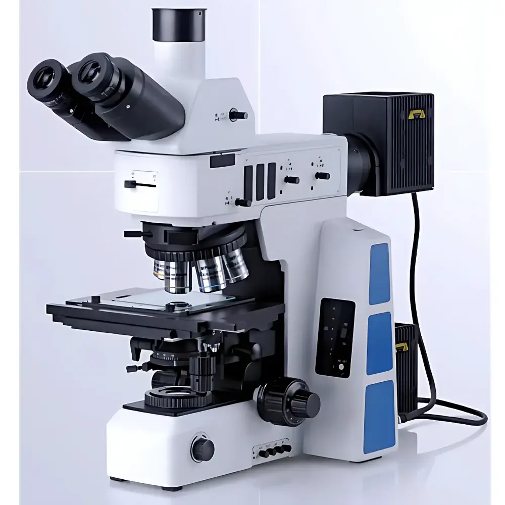
Lei-Tech LK-63M Research-Grade Upright Metallurgical Microscope
| Brand | Lei-Tech |
|---|---|
| Origin | Tianjin, China |
| Manufacturer Type | Direct Manufacturer |
| Product Type | Upright |
| Total Magnification | 50×–1000× |
| Eyepiece | PL10×/25 mm |
| Objectives | 5×, 10×, 20×, 50×, 100× (Infinity-Corrected, Semi-Apochromatic, Long Working Distance, Brightfield/Darkfield/Polarized/DIC-Compatible) |
| Optical System | Infinity-Corrected Dual-Path (UISC) |
| Illumination | 12 V / 100 W Halogen Lamp (Transmitted & Reflected, Pre-Centered) |
| Observation Modes | Brightfield, Darkfield, Polarized Light, Differential Interference Contrast (DIC) |
| Stage | Right-Handed 4-inch Mechanical Stage with Y-axis Lock & Glass Insert |
| Focus Mechanism | Coaxial Coarse/Fine Drive (25 mm Coarse Travel, 1 µm Fine Resolution) |
| Observation Tube | 30° Inclined, Trinocular, Infinity-Optimized, Pupil Distance 50–76 mm, Dual-Beam Splitting Ratio (100:0 or 0:100) |
| Condenser | Swing-Out Achromatic (NA 0.9) |
| Reflected Light Condenser | Brightfield/Darkfield Switchable, Adjustable Aperture & Field Diaphragms, Polarizer/Analyzer Slots, ND Filter Slot (ND50) |
| DIC Module | U-DICR Nomarski DIC System (Integrated into Objective Nosepiece) |
| Image Capture | 20 MP CMOS Sensor (5440 × 3648 px), Optional Integration |
Overview
The Lei-Tech LK-63M is a research-grade upright metallurgical microscope engineered for high-fidelity microstructural analysis of opaque, reflective specimens—primarily metals, alloys, ceramics, semiconductors, and advanced composites. It employs an infinity-corrected optical architecture (UISC — Universal Infinity Space Correction), enabling modular integration of contrast-enhancing components without compromising wavefront fidelity or axial alignment stability. Unlike finite-conjugate systems, the UISC design decouples magnification from tube length constraints, ensuring consistent resolution, chromatic correction, and illumination uniformity across all magnifications and observation modes. The system supports simultaneous and sequential use of brightfield (BF), darkfield (DF), polarized light (POL), and Nomarski differential interference contrast (DIC), making it suitable for both routine quality control and advanced failure analysis in materials science laboratories, semiconductor fabrication QA labs, and academic metallurgy departments.
Key Features
- Infinity-corrected semi-apochromatic long-working-distance (LWD) metallurgical objectives (5×–100×), optimized for BF/DF/POL/DIC with aluminum-alloy housings and multi-layer anti-reflective coatings for >92% transmission and minimal chromatic aberration.
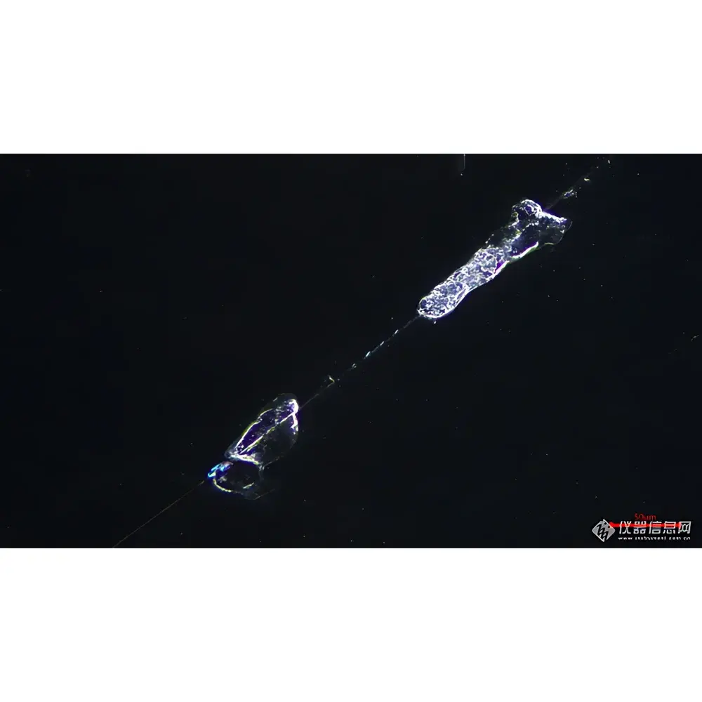
- Integrated U-DICR Nomarski DIC module—mounted directly at the nosepiece—enables quantitative height-difference visualization down to sub-100 nm surface topography, critical for evaluating scratch morphology, grain boundary relief, and thin-film stress patterns.
- Dual-path halogen illumination (12 V / 100 W) with pre-centered lamp housing, variable aperture and field diaphragms, and synchronized ND50 neutral density filter engagement during BF↔DF switching to prevent photostress and ensure observer comfort.
- Right-handed 4-inch mechanical stage with Y-axis locking mechanism, enabling precise linear scanning of wafer arrays or PCB substrates without positional drift; includes glass insert for optional transmitted-light applications (e.g., inclusion analysis in transparent phases).
- Trinocular observation head with 30° inclination, interpupillary adjustment (50–76 mm), and selectable beam-splitting ratio (100:0 or 0:100) for seamless integration with digital imaging systems compliant with USB 3.0 and GigE Vision standards.
- Coaxial coarse/fine focusing system with 25 mm vertical travel, 1 µm micrometer-driven fine adjustment, anti-slip tension control, and programmable upper limit stop—designed for repeatable Z-stack acquisition in automated metrology workflows.
Sample Compatibility & Compliance
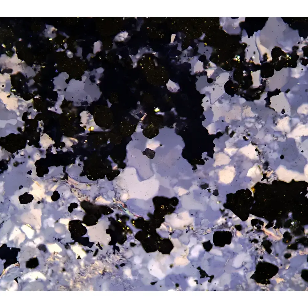
The LK-63M accommodates standard 25 mm and 32 mm diameter metallographic specimens (mounted or unmounted), as well as polished wafers up to 200 mm in diameter when used with optional large-stage adapters. Its dual-illumination path supports both reflected-light examination of conductive/non-conductive surfaces and transmitted-light inspection of semi-transparent phases (e.g., graphite nodules in ductile iron, oxide layers, or mineral inclusions). The system conforms to ISO 9001 manufacturing protocols and meets key optical performance benchmarks outlined in ASTM E3–22 (Standard Guide for Preparation of Metallographic Specimens) and ISO 4037-2 (Microscopy — Terminology for Optical Microscopes). All polarizing and DIC components are calibrated per ISO 10110-6 for retardation accuracy and extinction ratio (>1000:1). While not FDA 21 CFR Part 11–certified out-of-the-box, the microscope’s hardware architecture permits full audit-trail integration via third-party GLP/GMP-compliant image management software.
Software & Data Management
Though the base LK-63M configuration does not include embedded image analysis software, its trinocular port and standardized C-mount interface (23.2 mm flange distance) support plug-and-play integration with industry-standard platforms—including Olympus cellSens, Zeiss ZEN Blue, and open-source solutions such as FIJI/ImageJ. When paired with the optional 20 MP CMOS camera (5440 × 3648 px), the system enables high-resolution tiling (MIA), extended depth-of-field synthesis (EFI), and pixel-accurate measurement using NIST-traceable calibration slides. Raw TIFF output preserves bit-depth integrity for post-acquisition quantification of grain size (ASTM E112), phase fraction (ISO 13005), and line-profile roughness (ISO 4287). Metadata embedding (EXIF + custom XML tags) ensures traceability of magnification, illumination mode, objective ID, and stage coordinates—critical for ISO/IEC 17025–accredited testing laboratories.
Applications
- Semiconductor process control: Defect mapping on Si, GaN, and compound wafers using DIC-enhanced edge detection and polarization-sensitive void identification.
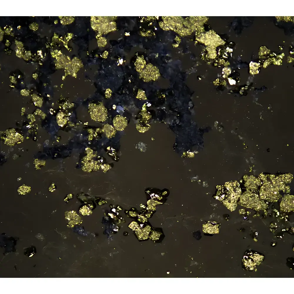
- Failure analysis in aerospace alloys: Grain boundary characterization in Ti-6Al-4V and nickel-based superalloys under thermal cycling stress, leveraging DF contrast for carbide precipitation and POL for twinning analysis.
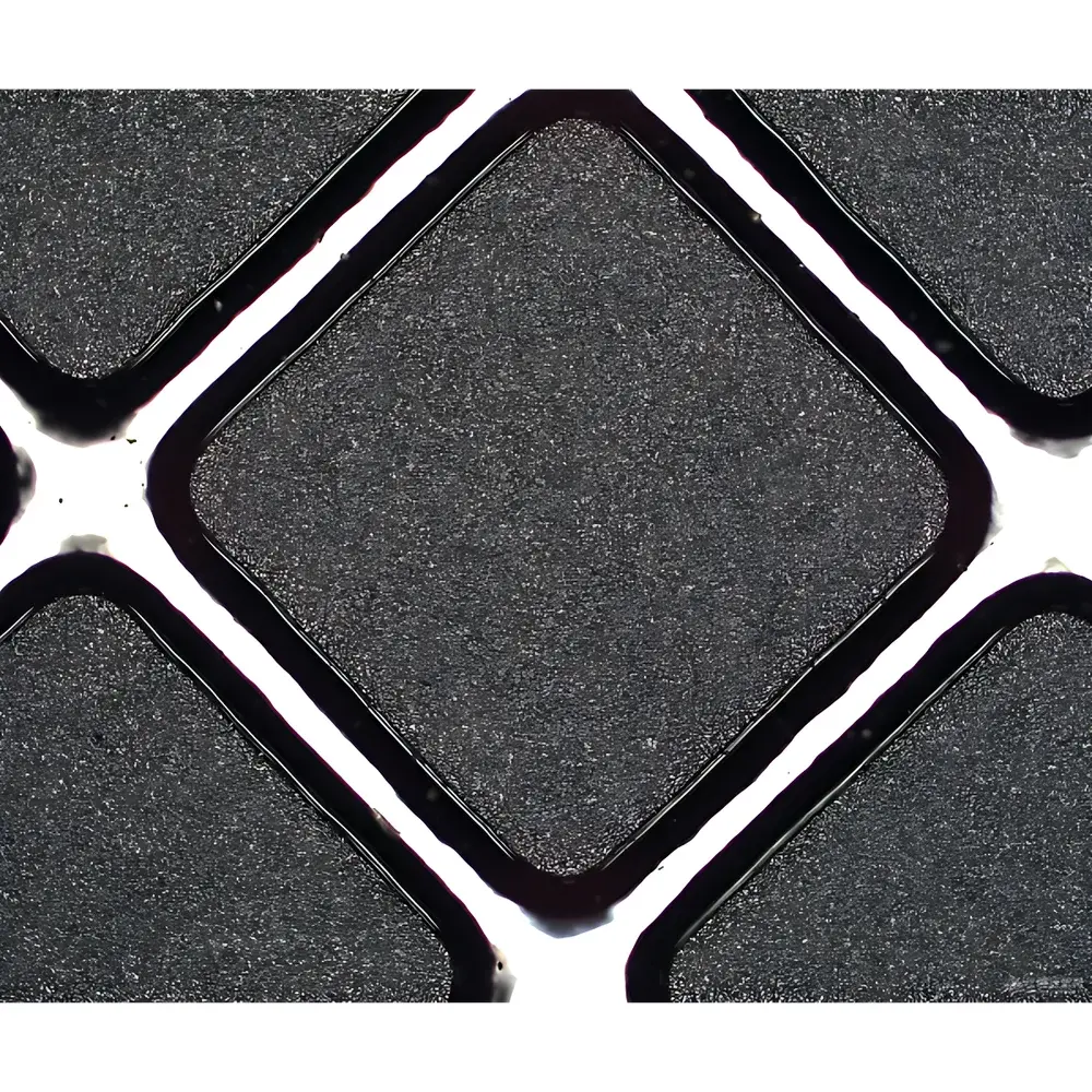
- Automotive cast iron evaluation: Quantitative assessment of graphite nodule count, shape factor, and matrix ferrite/pearlite ratio per ISO 945-1 and ASTM A247.
- Precision machining QA: Surface finish verification on hardened steel tooling using 100× DIC to resolve sub-micron feed marks and grinding burn signatures.
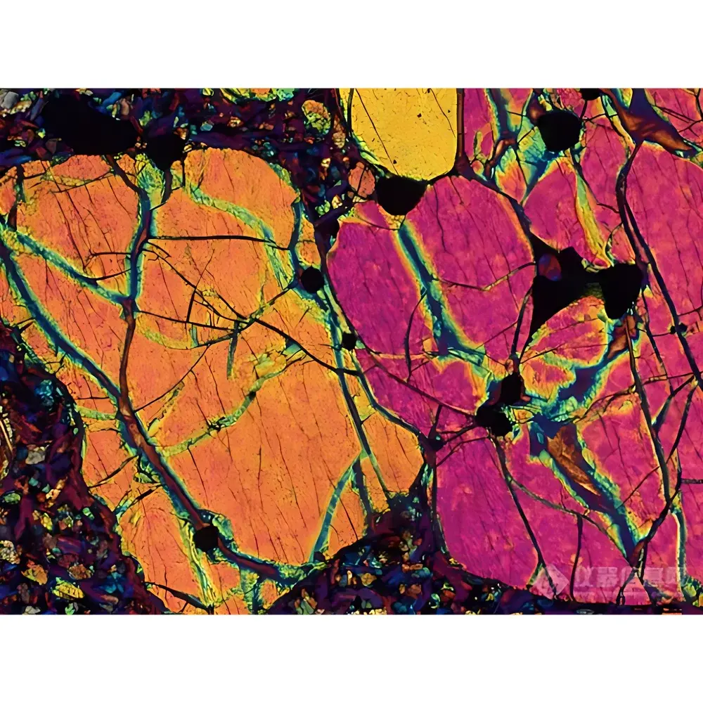
- Geological thin-section analysis: Birefringence mapping of quartz and feldspar in metamorphic rocks, enabled by 360° rotating analyzer and λ/4 compensator compatibility.
FAQ
Is the LK-63M compatible with fluorescence microscopy?
Yes—the semi-apochromatic LWD objectives transmit efficiently in the 365–650 nm range and support epi-fluorescence when fitted with appropriate excitation/emission filter cubes and a mercury or LED fluorescence illuminator (sold separately).
Can the microscope be upgraded to motorized focus or automated stage control?
Yes—its modular mechanical architecture accepts OEM-compatible stepper-motor kits for Z-axis automation and XY motorized stages with RS-232 or TTL trigger interfaces.
What is the warranty coverage and service response time?
Lei-Tech provides a 24-month limited warranty covering optical, mechanical, and electronic components. Technical support is available 7×24 via remote diagnostics; on-site service is dispatched within 72 hours for Tier-1 regions (North America, EU, APAC) following validated fault reporting.
Does the system meet ISO 10110 optical certification requirements?
All objectives and prisms undergo individual wavefront error testing (λ/10 PV @ 632.8 nm) and receive ISO 10110-3 compliance documentation upon request—valid for calibration traceability in accredited metrology environments.
How is parfocality maintained across BF, DF, and DIC modes?
The UISC optical pathway, combined with precision-machined DIC prism mounts and objective-specific correction collars, ensures <±2 µm axial deviation across all five objectives and all contrast modes—verified per ISO 8578 Annex B.


