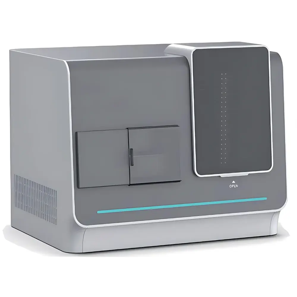
LEI-TECH LK-PowerScin Series Fully Automated Digital Pathology Scanner
| Brand | LEI-TECH |
|---|---|
| Origin | Tianjin, China |
| Manufacturer Type | OEM Manufacturer |
| Product Origin | Domestic (China) |
| Model | LK-PowerScinP3 / LK-PowerScinP30/60/120 |
| Pricing | Available Upon Request |
Overview
The LEI-TECH LK-PowerScin Series is a fully automated digital pathology scanner engineered for high-fidelity, high-throughput whole-slide imaging (WSI) in clinical pathology, translational research, and diagnostic laboratories. Based on continuous push-broom scanning with ultra-short exposure time and adaptive real-time autofocus, the system captures sub-micron-resolution optical images across standard 25 mm × 75 mm glass slides without mechanical stitching artifacts. Its core optical architecture features a large-aperture, low-distortion lens design optimized for 20× and optional 40× objective magnifications, coupled with a high-dynamic-range magnetic-levitation focus actuator enabling precise Z-axis control within ±0.1 µm repeatability. The scanner operates on a dual-layer heterogeneous stage driven by linear magnetic motors, ensuring synchronized X-Y-Z motion with closed-loop positional feedback. Unlike conventional step-and-repeat scanners, the LK-PowerScin employs continuous-area scanning—reducing acquisition time while preserving image fidelity, signal-to-noise ratio, and spatial coherence across the entire field of view.
Key Features
- Real-time adaptive autofocus: Continuous depth-of-field tracking during scanning ensures consistent sharpness across uneven tissue topographies and varying section thicknesses (e.g., FFPE, cytology smears, IHC-stained slides).
- High-speed acquisition: Full-scan time ≤50 seconds at 20× (15 mm × 15 mm ROI), ≤80 seconds at 40×—achievable via short-exposure push-broom illumination and synchronized line-sensor readout.
- Precision motion control: XYZ axes with 0.1 µm resolution and full closed-loop feedback; dual-layer heterostructure stage minimizes vibration-induced blur and thermal drift.
- Flexible slide handling: LK-PowerScinP3 supports up to 3 standard slides per batch; LK-PowerScinP30/60/120 models accommodate 30, 60, or 120 slides with auto-loading, barcode recognition, and priority-based emergency insertion.
- Optical integrity: Proprietary low-aberration optics calibrated for visible-light spectral range (400–700 nm); compatible with brightfield, immunohistochemistry (IHC), and chromogenic in situ hybridization (CISH) workflows.
- Robust software integration: Native support for DICOM-SR, OpenSlide, and ASPIRE-compliant metadata embedding; compliant with CAP and CLIA pre-analytical documentation requirements.
Sample Compatibility & Compliance
The LK-PowerScin series is validated for routine diagnostic-grade imaging of formalin-fixed paraffin-embedded (FFPE) tissue sections, liquid-based cytology (LBC) preparations, blood smears, tissue microarrays (TMAs), and fluorescence-labeled specimens (with appropriate filter sets). All scanning protocols adhere to ISO 15189:2022 Annex B guidelines for digital pathology validation, including focus stability testing, color fidelity assessment (Delta E ≤3.0), and geometric distortion verification (<0.1% across FOV). The system supports GLP/GMP-aligned audit trails, user authentication, and FDA 21 CFR Part 11–compliant electronic signatures when deployed with certified LIS/PACS interfaces. Regulatory documentation—including CE marking under IVDR Class C (pending), NMPA registration (Class II), and ISO 13485:2016 QMS certification—is available upon request.
Software & Data Management
The bundled LEI-SCIN Software Suite provides end-to-end WSI workflow management: automatic slide detection, region-of-interest (ROI) segmentation, multi-plane focus stacking, seamless tile stitching, and lossless compression (TIFF, SVS, NDPI export options). It includes DICOM WSI conformance (Supplement 145), embedded annotation layers, synchronized multi-user viewing, and RESTful API access for integration into enterprise PACS, telepathology platforms, or AI training pipelines. Cloud upload functionality supports encrypted TLS 1.3 transmission and HIPAA-compliant storage architectures. Audit logs record every scan event—including operator ID, timestamp, objective used, focus map, and QC flags—with immutable retention configurable per institutional policy.
Applications
- Clinical diagnostics: Routine histopathology digitization for primary diagnosis, second opinion, and remote consultation—validated for cervical cancer screening (ASC-US/LSIL/HSIL classification) with AI-assisted triage achieving >60% negative predictive value.
- Research pathology: High-content analysis of TMAs and spatial biomarker mapping; integration-ready with QuPath, HALO, and Visiopharm modules.
- Cytogenetics: Chromosome karyotyping via high-resolution 40× scanning; supports automated centromere/telomere detection and aberration classification using reference databases containing >10,000 annotated abnormal karyotypes.
- Quality assurance: Digital slide archiving with zero degradation over time; enables longitudinal comparison, inter-laboratory proficiency testing, and CAP-accredited slide review cycles.
- Education & training: Real-time collaborative annotation, case-based teaching libraries, and mobile-responsive web viewer for tablets and desktops—fully responsive across iOS, Android, and Windows environments.
FAQ
Does the LK-PowerScin support both brightfield and fluorescence imaging?
Yes—brightfield imaging is standard; fluorescence capability requires optional LED excitation modules and emission filter wheels (available as add-on kits).
What file formats are natively supported for export and archival?
TIFF (uncompressed and LZW-compressed), SVS (Aperio), NDPI (Hamamatsu), and DICOM WSI (Suppl. 145) are fully supported; metadata conforms to OpenSlide and ASPIRE standards.
Is the system compatible with existing LIS/PACS infrastructure?
Yes—the scanner integrates via HL7 v2.x, DICOM WSI, and REST APIs; HL7 ORM/OBR message handling and DICOM Modality Worklist (MWL) are enabled by default.
How is focus accuracy validated across large tissue sections?
Each scan includes dynamic focus mapping using contrast-gradient optimization; Z-stack fusion is applied where tissue thickness exceeds depth-of-field, with QC reports generated per slide.
What cybersecurity measures are implemented for data-in-transit and data-at-rest?
All network communications use TLS 1.3 encryption; locally stored images may be encrypted using AES-256; role-based access control (RBAC) and session timeout policies align with NIST SP 800-53 Rev. 5 controls.


