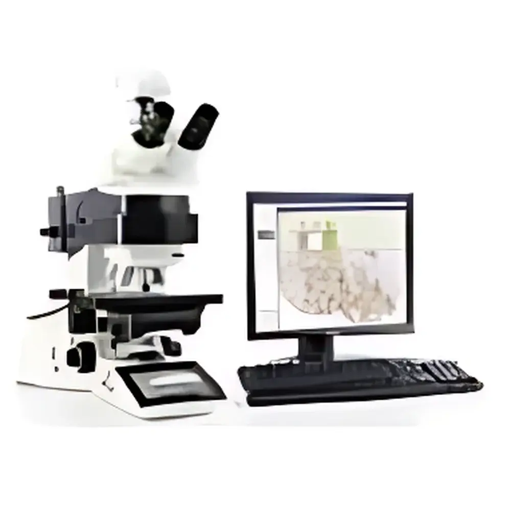
Leica Aperio Ariol Fluorescence In Situ Hybridization (FISH) Slide Scanner
| Brand | Leica |
|---|---|
| Origin | Germany |
| Model | Aperio Ariol |
| Slide Capacity | 4, 8, or 200 slides (with SL200 loader) |
| Fluorescence Channels | Up to 9 |
| Z-Stack Layers | Up to 30 |
| Clinical Validation | CE-IVD and FDA-cleared analysis modules available |
| Integration | Compatible with Aperio eSlide Manager and DIH (Digital Imaging Hub) |
| Microscope Platform | Leica DM600B automated upright microscope |
| Oil Immersion | Automated oil dispensing system |
| Software | Aperio Image Analysis Suite with real-time algorithm parameter tuning |
Overview
The Leica Aperio Ariol Fluorescence In Situ Hybridization (FISH) Slide Scanner is a high-throughput, clinical-grade whole-slide imaging (WSI) platform engineered for quantitative fluorescence cytogenetic analysis. Built upon the Leica DM600B fully automated upright microscope architecture, the Ariol system combines precision optical design with robust motorized stage control, multi-channel fluorescence excitation/emission filtering, and programmable Z-stack acquisition to deliver reproducible, high-fidelity digital FISH images. Unlike conventional brightfield-only scanners, the Ariol integrates dual-modality imaging—supporting both brightfield and high-sensitivity fluorescence scanning within a single, unified hardware and software environment. Its core function is to digitize FISH-stained tissue sections and cytology preparations at clinically validated resolutions (typically 20× or 40× objective magnification), enabling downstream quantitative signal enumeration, spatial localization, and pattern recognition in accordance with standardized cytogenetic interpretation guidelines.
Key Features
- Multi-channel fluorescence acquisition supporting up to 9 independent excitation/emission filter sets—enabling simultaneous detection of multiple FISH probes (e.g., HER2, EGFR, ALK, CEP17, centromeric, and locus-specific probes) without spectral crosstalk.
- Programmable Z-stack imaging with up to 30 focal planes per field-of-view and user-defined step size—critical for accurate signal counting in thick or unevenly hybridized tissue sections.
- Flexible slide handling: supports manual loading (4- or 8-slide trays) or high-capacity automated loading via the optional SL200 robotic slide loader (200-slide capacity), enabling unattended overnight scanning.
- Integrated automated oil immersion delivery system ensures consistent refractive index matching across large-area fluorescence acquisitions—minimizing spherical aberration and signal loss at high numerical aperture (NA).
- Real-time image analysis parameter adjustment within the Aperio Image Analysis Suite—allowing pathologists and cytogeneticists to optimize segmentation, thresholding, and spot-counting algorithms directly on acquired data before final quantification.
- Compliance-ready architecture: supports audit trails, user authentication, and electronic signatures in alignment with CLIA, CAP, ISO 15189, and FDA 21 CFR Part 11 requirements when deployed with validated configurations.
Sample Compatibility & Compliance
The Ariol system accommodates standard 1″ × 3″ glass microscope slides with coverslips (thickness 0.13–0.17 mm), including formalin-fixed paraffin-embedded (FFPE) tissue sections, touch preps, bone marrow aspirates, and interphase/metaphase cytology preparations. It supports common FISH probe chemistries (directly labeled fluorophores, hapten-based indirect detection) and hybridization protocols from major vendors (Abbott Molecular, Agilent, Dako/Agilent, Cytocell). All analytical modules used in routine clinical reporting—including HER2 amplification scoring, ALK rearrangement detection, and EGFR copy number assessment—have received CE-IVD marking in Europe and FDA 510(k) clearance in the United States. The platform conforms to ISO/IEC 17025 and ISO 13485 quality management standards for in vitro diagnostic medical devices.
Software & Data Management
Acquired FISH images are natively stored in Aperio SVS format (multi-resolution pyramidal TIFF), compatible with PACS, LIS, and enterprise imaging systems via DICOM-SR and HL7 interfaces. Integration with Aperio eSlide Manager enables centralized slide metadata indexing, role-based access control, and secure web-based review across geographically distributed laboratories. The Digital Imaging Hub (DIH) facilitates seamless workflow orchestration—linking scanning, analysis, reporting, and archival functions while maintaining full traceability of image processing history. All analysis sessions generate immutable audit logs recording operator ID, timestamp, parameter values, and versioned algorithm binaries—supporting GLP/GMP and regulatory inspection readiness.
Applications
The Ariol platform serves as a primary digital cytogenetics tool in academic medical centers, reference laboratories, and pharmaceutical biomarker development units. Key use cases include: quantitative HER2 gene amplification assessment in breast cancer biopsies per ASCO/CAP guidelines; ALK fusion detection in non-small cell lung carcinoma using break-apart probe assays; EGFR copy number variation analysis in glioblastoma and head-and-neck squamous cell carcinoma; and polysomy/aneuploidy evaluation in hematologic malignancies. Its ability to generate permanent, shareable digital records supports remote expert consultation, longitudinal case review, and retrospective clinical trial biomarker re-evaluation.
FAQ
Does the Ariol support both brightfield and fluorescence scanning on the same slide?
Yes—the system acquires brightfield and fluorescence images sequentially or concurrently using dedicated illumination paths and filter wheels, enabling correlative morphology and probe signal analysis.
Can Z-stack parameters be defined per tissue region or only globally per slide?
Z-stack settings are configurable per scan region of interest (ROI), allowing optimization for heterogeneous samples such as tumor-infiltrated stroma or layered epithelial structures.
Is the SL200 loader compatible with all Ariol configurations?
The SL200 robotic loader requires the Ariol XL configuration and integration with Leica’s DM600B microscope base equipped with extended travel stage and reinforced frame.
Are analysis algorithms validated for clinical reporting in the U.S.?
Yes—multiple FISH analysis modules have FDA 510(k) clearance for use in clinical diagnostics, with documented sensitivity, specificity, and inter-reader concordance metrics per CLSI EP12-A2 guidelines.
How does the system ensure focus stability during long-duration overnight scans?
Ariol employs continuous autofocus using infrared-based contrast detection and adaptive Z-tracking—compensating for thermal drift and mechanical relaxation over multi-hour acquisitions.


