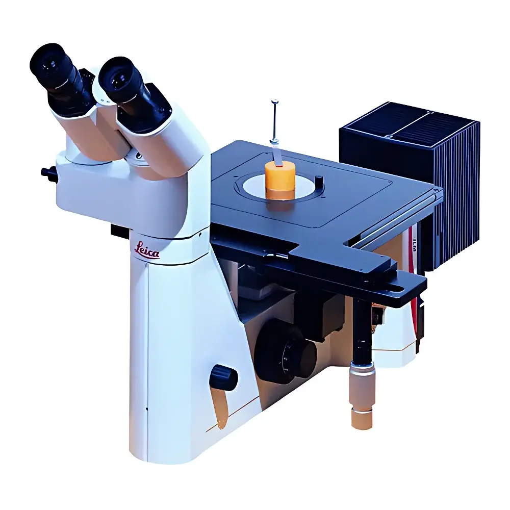
Leica DM ILM Inverted Metallurgical Microscope
| Brand | Leica |
|---|---|
| Origin | Germany |
| Model | DM ILM |
| Configuration | Inverted |
| Imaging System | Integrated Digital Image Analysis Ready |
| Total Magnification Range | 50× to 1000× |
| Eyepieces | 10× (standard), optional 12.5×, 16×, 25× (18–20 mm field of view) |
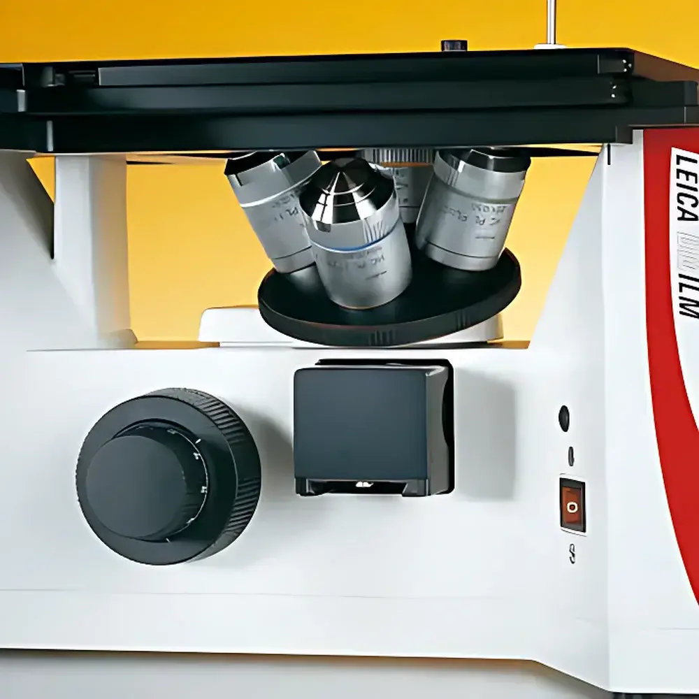
| Objectives | 1.25×, 5×, 10×, 20×, 50×, 100×, 250× |
| Objective Turret | 4-position, M25 thread, brightfield-optimized |
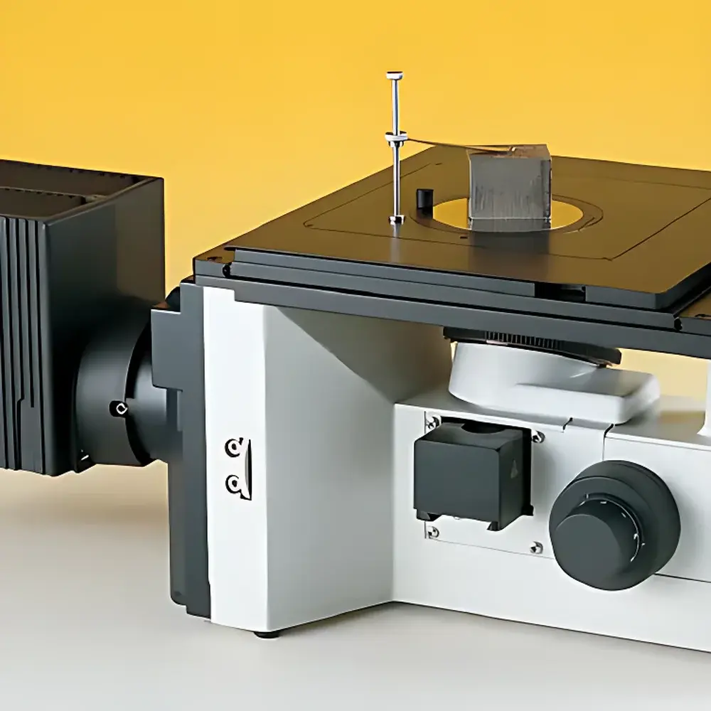
| Stage Dimensions | 247 × 230 mm, XY travel 60 × 40 mm |
| Focus Mechanism | Coaxial coarse/fine adjustment (±7 mm upward / −2 mm downward) |
| Illumination | LED cold-light source (≥20,000 h lifetime), insertable filter sets including polarizing components (polarizer & analyzer) |
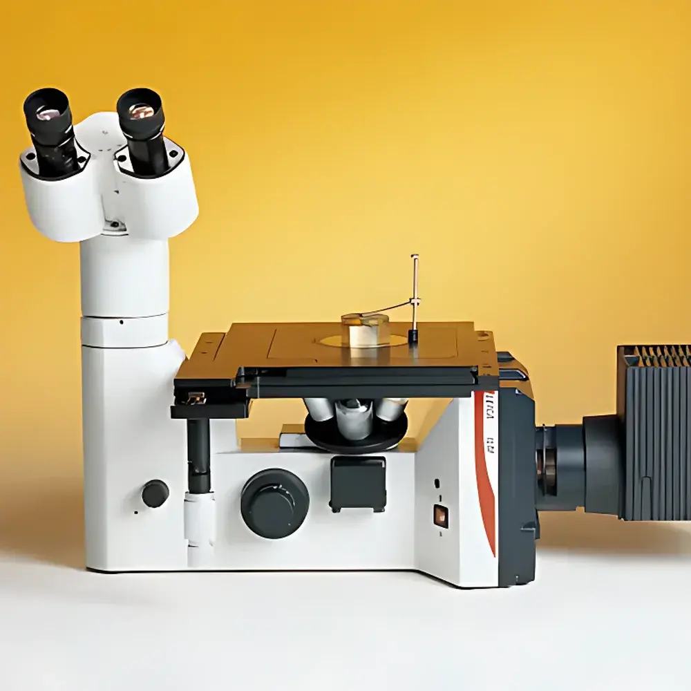
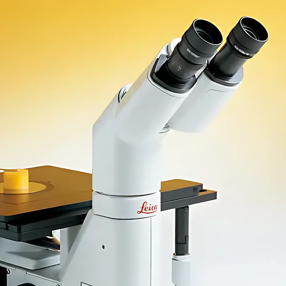
| Viewing Head | 30° inclined binocular tube, interpupillary adjustment 48–75 mm, side-port 100% beam split for imaging |
| Optical Design | HC (High Contrast) infinity-corrected optical system with axial and radial chromatic aberration correction |
Overview
The Leica DM ILM Inverted Metallurgical Microscope is an engineered solution for high-precision microstructural analysis of opaque, polished, and etched metallic and industrial material specimens. Designed specifically for metallurgy laboratories, quality control facilities, and failure analysis centers, the DM ILM employs a robust inverted configuration—placing the objective lenses beneath the specimen stage—to accommodate large, heavy, or irregularly shaped samples without compromising optical access or mechanical stability. Its core optical architecture utilizes Leica’s proprietary HC (High Contrast) infinity-corrected optics, featuring simultaneous axial and radial chromatic aberration correction across the full magnification range. This optical design eliminates stray light artifacts, enhances edge definition, and delivers consistent contrast and resolution from low-magnification overview imaging (50×) to high-resolution microstructural evaluation (1000×). The system supports standardized observation modes including brightfield and polarized light—enabling crystallographic orientation analysis, phase identification, inclusion characterization, and grain boundary delineation in accordance with ASTM E3, ISO 643, and USP metallographic evaluation guidelines.
Key Features
- Inverted platform with fixed stage and motorized or manual objective turret—ideal for bulk specimens, mounted cross-sections, and hot-stage or environmental chamber integration.
- HC infinity-corrected optical path optimized for uniform flat-field performance, minimal distortion, and high transmission efficiency across visible wavelengths (400–700 nm).
- 4-position M25-thread objective turret pre-aligned for brightfield use; compatible with Leica’s full suite of metallurgical objectives—including long-working-distance, strain-free, and DIC-capable variants.
- Stable mechanical stage (247 × 230 mm) with coaxial X–Y controls and 60 × 40 mm travel range; accommodates standard petri dishes, wafer carriers, and custom fixtures via interchangeable aperture plates.
- Dedicated LED illumination module delivering stable, flicker-free, color-temperature-consistent output (5700 K), with integrated neutral density and interference filters for quantitative intensity control.
- Quick-switch polarizing optics: slide-in polarizer and analyzer units enable single-hand activation of polarized contrast—critical for birefringent phase detection in alloys, ceramics, and composites.
- Ergonomic 30° inclined binocular head with 48–75 mm interpupillary adjustment and 100% light-splitting side port for synchronized live imaging and visual inspection.
Sample Compatibility & Compliance
The DM ILM accepts standard metallographic specimens up to 50 mm in diameter and 40 mm in height, including mounted epoxy or phenolic resin blocks, as-received castings, rolled sheets, weld zones, and additive-manufactured parts. Its inverted geometry permits direct mounting on standard laboratory tables or integration into automated sample-handling systems. All optical and mechanical components comply with DIN EN ISO 9001 manufacturing standards. The system supports GLP/GMP workflows through traceable calibration protocols (per ISO/IEC 17025), and when paired with Leica Application Suite (LAS) X software, meets audit-trail and electronic signature requirements under FDA 21 CFR Part 11 for regulated environments.
Software & Data Management
The microscope is fully compatible with Leica LAS X software—a modular, CE-IVD and FDA-listed platform supporting image acquisition, measurement, annotation, and report generation. Optional modules include Extended Depth of Field (EDF) for focus stacking of rough or tilted surfaces, Quantitative Metallography (QMET) for grain size (ASTM E112), inclusion rating (ASTM E45), and phase fraction analysis, and Automated Particle Analysis (APA) for size distribution and morphology metrics. Raw image data is stored in TIFF or Leica’s proprietary LIF format, preserving metadata (objective ID, magnification, exposure time, filter status) for full experimental reproducibility.
Applications
- Metallographic examination of ferrous and non-ferrous alloys per ASTM E3, ISO 643, and JIS G 0551.
- Weld microstructure assessment—including heat-affected zone (HAZ) characterization, porosity quantification, and intermetallic phase mapping.
- Failure analysis of fracture surfaces, fatigue cracks, and stress-corrosion cracking features.
- Quality assurance of sintered powders, thermal spray coatings, and composite laminates.
- Research-grade microstructural evolution studies during heat treatment, creep testing, or deformation experiments.
- Education and training in materials science curricula requiring standardized, repeatable imaging protocols.
FAQ
What is the maximum usable magnification for quantitative metallographic analysis?
For statistically valid grain size or phase fraction measurements per ASTM E112, the recommended upper limit is 500× using a 50× objective and 10× eyepiece. Higher magnifications (up to 1000×) are suitable for qualitative defect identification but require careful depth-of-field management.
Can the DM ILM be upgraded for differential interference contrast (DIC)?
Yes—DIC functionality can be added via Leica’s optional Wollaston prism sets and Nomarski-compatible objectives, enabling nanoscale topographic contrast without staining.
Is the LED illumination intensity adjustable and calibrated?
Intensity is continuously variable via software or front-panel control and traceably calibrated against NIST-traceable photometric standards using Leica’s optional light meter accessory.
Does the system support motorized Z-axis focus for automated layer scanning?
Motorized focus is available as an OEM option (Leica Z Drive) and integrates seamlessly with LAS X for multi-layer focus stacking, tilt-series acquisition, and serial sectioning workflows.
How is optical alignment verified and maintained?
Leica provides factory-certified alignment reports and includes built-in centering tools for Köhler illumination setup; routine verification follows ISO 10934-1 procedures using certified test targets.


