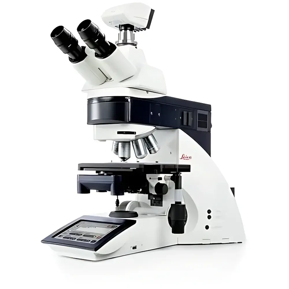
Leica DM5500 B Automated Upright Microscopy System for Life Science Research
| Brand | Leica |
|---|---|
| Origin | Germany |
| Model | DM5500 B |
Overview
The Leica DM5500 B is an automated upright microscope system engineered specifically for demanding life science applications—including live-cell imaging, fluorescence in situ hybridization (FISH), immunofluorescence assays with deconvolution, and high-precision morphological analysis. Built upon Leica’s legacy of optical excellence, the DM5500 B implements a robust mechanical architecture combined with precise motorized actuation and encoded optical components to deliver reproducible, quantitative imaging under multiple contrast modalities. Its core optical design follows Köhler illumination principles and integrates advanced beam path management for simultaneous optimization across brightfield, phase contrast, differential interference contrast (DIC), and epifluorescence channels. The system operates on a rigid, vibration-damped baseplate and features thermal-stable optics housing—critical for maintaining focus stability during time-lapse experiments lasting several hours.
Key Features
- Motorized Z-axis drive with sub-micron repeatability, enabling automated refocusing, multi-plane z-stack acquisition, and memory-based focus recall across objectives.
- Encoded 7-position objective turret ensures automatic recognition and calibration of magnification, numerical aperture, and correction collar settings—eliminating manual reconfiguration between objectives.
- World’s first fully automated DIC system: Upon selection of objective and contrast mode, the microscope autonomously inserts and aligns Wollaston prisms, polarizers, and analyzers into the optical path, optimizing shear direction and bias retardation per objective for consistent contrast and quantitative birefringence assessment.
- Integrated SmartTouch touchscreen interface provides direct hardware-level control of illumination intensity, filter cube selection, stage movement, and focus presets—all synchronized with LAS and LAS AF software environments.
- CCIC (Constant Color and Intensity Control) dynamically adjusts halogen lamp voltage and neutral density filtering to maintain stable color temperature (≈3200 K) and luminous flux across varying brightness levels—essential for comparative photometric analysis and long-term exposure consistency.
- Apochromatically corrected fluorescence optics minimize axial chromatic shift and spherical aberration, supporting high-resolution multicolor imaging with minimal crosstalk—even at 100× oil immersion.
Sample Compatibility & Compliance
The DM5500 B accommodates standard microscopy formats including glass slides (26 × 76 mm), petri dishes (35–100 mm), multi-well plates (6–96-well), and custom chambered coverslips. Its modular stage design supports motorized XY translation with optional environmental control integration (temperature, CO₂, humidity). The system conforms to IEC 61000-6-3 (EMC emissions) and IEC 61000-6-2 (immunity), and meets CE marking requirements for laboratory instrumentation. While not inherently 21 CFR Part 11 compliant, LAS AF software offers optional audit trail, electronic signature, and user access control modules suitable for GLP/GMP-aligned workflows when deployed with validated IT infrastructure.
Software & Data Management
Control and acquisition are managed via Leica Application Suite (LAS) and LAS Advanced Fluorescence (LAS AF), both supporting TIFF, JPEG2000, and native .lei file formats. LAS AF enables multi-dimensional acquisition (x-y-z-t-λ), spectral unmixing, region-of-interest (ROI)-based intensity quantification, and batch processing of deconvolution algorithms (e.g., constrained iterative deconvolution). Metadata embedding includes objective ID, DIC prism position, exposure time, gain, lamp intensity, and environmental sensor readings (when interfaced). Export options support MIAME-compliant annotation and integration with OMERO or ImageJ/Fiji pipelines via open API protocols.
Applications
- Live-cell dynamics: Time-lapse DIC and phase contrast imaging of mitosis, organelle trafficking, and cytoskeletal remodeling without phototoxicity.
- FISH and chromosomal mapping: High-contrast, low-background fluorescence detection with automated focus maintenance across large tissue sections.
- Immunofluorescence quantification: Multi-channel co-localization analysis supported by apochromatic optics and hardware-synchronized filter switching.
- Neuroscience preparations: Imaging of acute brain slices using infrared-DIC (IR-DIC) compatibility and adjustable condenser alignment for thick specimen visualization.
- Developmental biology: Long-duration embryo imaging with thermal stabilization and automated z-series acquisition synchronized to developmental milestones.
FAQ
Does the DM5500 B support objective correction collar adjustment automation?
Yes—the encoded turret communicates objective-specific correction collar parameters to the system firmware, enabling motorized collar positioning when switching between objectives with differing cover slip thickness requirements.
Can third-party cameras be integrated with LAS AF software?
LAS AF supports DCAM-compliant IEEE 1394 and USB3 Vision cameras; full driver certification is required for guaranteed synchronization with hardware-triggered acquisition modes.
Is DIC alignment saved per objective or per magnification?
DIC alignment parameters—including prism centering, shear axis orientation, and bias offset—are stored per objective ID, ensuring optimal configuration regardless of zoom or intermediate magnification changes.
What is the maximum z-stack depth achievable with motorized focus?
With standard 10 mm travel range and optional extended-focus modules, z-stacks up to 1000 µm can be acquired with ≤10 nm step resolution, limited only by objective working distance and sample transparency.
How does CCIC affect quantitative fluorescence measurements?
CCIC stabilizes photon flux and spectral output over time, reducing drift-related artifacts in intensity-based assays—particularly critical for ratiometric or kinetic measurements spanning >30 minutes.


