Leica FL560 Fluorescence Module for Neurosurgical Microscopes
| Brand | Leica |
|---|---|
| Origin | Germany |
| Manufacturing Region | Europe |
| Module Type | Integrated Surgical Fluorescence Imaging Module |
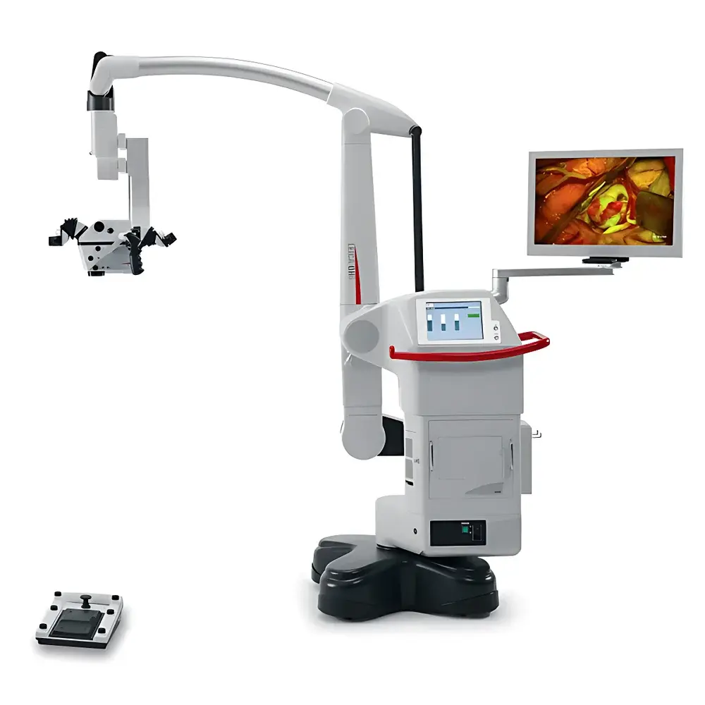
| Compatible Microscope Platforms | Leica M530 OH5, M720 OH5 |
| Excitation Range | 460–500 nm (Blue) |
| Emission Detection Range | >510 nm (Green–Yellow–Red Spectrum) |
| Optical Architecture | Dual-Channel Simultaneous White Light + Fluorescence Imaging |
| Integration Standard | Leica OpenArchitecture™ Platform |
| Compliance | Designed for ISO 13485-certified neurosurgical environments |
| Regulatory Context | Supports GLP/GMP-aligned documentation workflows when paired with Leica LAS X software and FDA 21 CFR Part 11-compliant acquisition systems |
Overview
The Leica FL560 Fluorescence Module is an integrated optical imaging component engineered for high-precision intraoperative fluorescence visualization in neurosurgical microscopes. It operates on the principle of narrow-band excitation and spectral separation—utilizing optimized dichroic filters and bandpass emission optics to isolate fluorescent signal from ambient white light illumination in real time. Unlike sequential or alternating-mode fluorescence systems, the FL560 enables true simultaneous dual-channel imaging: anatomical detail under standard white-light illumination is preserved at full resolution while fluorophore-specific emission (>510 nm) is captured in parallel without temporal lag or registration drift. This capability is critical in microsurgical contexts where spatial fidelity between structural landmarks and functional fluorescence signals—such as tumor margins, vascular perfusion, or cortical activation—must be maintained at sub-millimeter accuracy. The module is optically calibrated to leverage Leica’s apochromatic microscope objectives and color-corrected beam paths, ensuring minimal chromatic aberration across both visible and extended green–yellow emission bands.
Key Features
- Simultaneous white-light and fluorescence imaging with zero frame latency or image shift
- Optimized excitation filter set centered at 480 nm ±10 nm, compatible with clinically validated fluorophores including fluorescein sodium, 5-ALA-induced PpIX, and indocyanine green (ICG) under blue-light excitation protocols
- Dual-path optical design with dedicated emission pathways for >510 nm signal capture, minimizing bleed-through and maximizing signal-to-noise ratio
- Seamless integration via Leica OpenArchitecture™—no external light sources, cameras, or third-party controllers required
- Single-button activation via ergonomic hand controller or footswitch; mode switching between FL560, FL400*, and FL800 (on TriFluoro-enabled M530 platforms) is hardware-synchronized
- Native support for Leica LAS X navigation software, enabling synchronized timestamping, metadata tagging (e.g., exposure time, gain, illumination intensity), and DICOM-SR export for PACS integration
Sample Compatibility & Compliance
The FL560 module is validated for use with fluorophores exhibiting peak excitation between 460 nm and 500 nm and emission maxima above 510 nm—including but not limited to sodium fluorescein (Ex/Em: 494/521 nm), protoporphyrin IX (PpIX) (Ex/Em: 405–420/635–710 nm, with secondary blue-excitation response), and ICG under off-label blue-light protocols. It complies with IEC 60601-2-57 for medical electrical equipment safety and meets optical performance criteria outlined in ISO 10940:2021 (Microscopes — Requirements for surgical microscopes). When deployed in regulated clinical environments, the system supports audit-trail generation, user-access controls, and electronic signature workflows when operated with Leica LAS X software configured per FDA 21 CFR Part 11 requirements.
Software & Data Management
Leica LAS X software provides unified control of illumination, camera parameters, and recording settings for both white-light and FL560 modes. Each mode retains independent calibration profiles: automatic gain adjustment, dynamic range optimization, and gamma correction are applied contextually based on real-time histogram analysis. Video streams are encoded in H.264 at up to 60 fps (1080p) with embedded SMPTE timecode. All recordings include EXIF-style metadata—microscope model, objective magnification, FL mode active, exposure duration, and DICOM-compatible patient anonymization fields. Export formats include AVI, MP4, and DICOM-RT structure sets for longitudinal case review and multicenter trial repositories.
Applications
- Intraoperative delineation of glioblastoma resection margins using 5-ALA fluorescence
- Real-time assessment of cortical perfusion during aneurysm clipping or bypass surgery
- Identification of parathyroid glands in thyroidectomy via autofluorescence enhancement
- Functional mapping of eloquent cortex using intrinsic signal imaging protocols
- Training and simulation: HD video archives enable standardized assessment of surgical decision-making under fluorescence guidance
FAQ
Is the FL560 module compatible with non-Leica microscopes?
No. The FL560 is mechanically, optically, and electronically designed exclusively for integration with Leica M530 OH5 and M720 OH5 surgical microscope platforms.
Does FL560 require external excitation light sources or cameras?
No. It utilizes the microscope’s built-in LED illumination engine and native CMOS sensor array—no additional hardware installation is necessary.
Can FL560 be retrofitted onto older Leica microscope models?
Only M530 OH5 and M720 OH5 units manufactured after Q3 2018 with OpenArchitecture™ firmware v3.2 or later support FL560 integration. Earlier platforms lack the optical rail interface and control bus architecture.
What regulatory documentation is provided for clinical validation?
Leica supplies a Technical File compliant with EU MDR Annex II, including optical performance test reports, biocompatibility data (ISO 10993-5), and electromagnetic compatibility (EMC) test certificates per EN 60601-1-2.
How is fluorescence quantification handled?
LAS X supports region-of-interest (ROI) intensity profiling and relative fluorescence unit (RFU) calculation against calibrated reference standards; absolute quantification requires external photometric calibration using NIST-traceable reference fluorophores.


