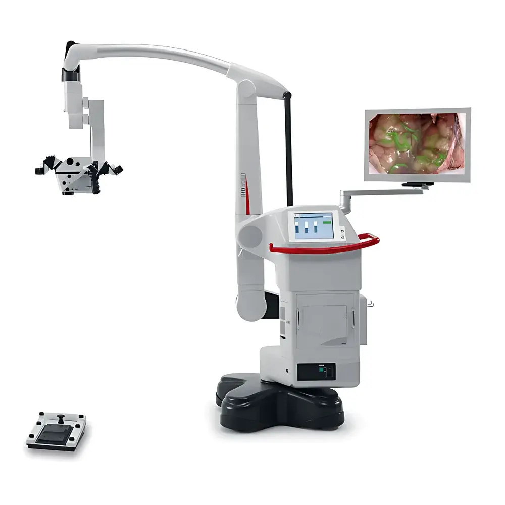
Leica GLOW800 Surgical Microscope for Intraoperative Fluorescence Imaging
| Brand | Leica |
|---|---|
| Country of Origin | Germany |
| Model | GLOW800 |
| Instrument Type | Stereoscopic Fluorescence Surgical Microscope |
| Excitation Source | High-Power LED |
| Integration Platform | Fully Integrated with Leica M530 Surgical Microscope |
| Display | Full HD 1080p CaptiView In-Eyepiece Projection System |
| Contrast Ratio | 500:1 |
| Color Rendering | Multi-spectral AR Fusion (Natural Tissue + Pseudocolor ICG Fluorescence) |
| Optional Module | 3D Visualization & Recording |
Overview
The Leica GLOW800 is a purpose-engineered intraoperative fluorescence imaging module designed exclusively for integration with the Leica M530 surgical microscope platform. It leverages advanced multi-spectral optical architecture and real-time image fusion algorithms to overlay indocyanine green (ICG) fluorescence signals—captured in the near-infrared (NIR) range—onto high-fidelity, color-accurate white-light stereoscopic views. Unlike conventional monochrome NIR fluorescence systems that require mode switching and mental registration between anatomical and vascular datasets, the GLOW800 delivers a single, spatially registered, depth-preserving augmented reality (AR) view. This capability is grounded in dual-path optical beam splitting, synchronized LED excitation (785 nm peak), and proprietary spectral deconvolution processing, enabling simultaneous visualization of microvascular perfusion dynamics and fine neuroanatomical landmarks without perceptual latency or ocular refocusing.
Key Features
- Fully integrated AR fluorescence mode—activated via ergonomic hand control or footswitch on the M530 platform
- CaptiView in-eyepiece projection system delivering full HD 1080p AR overlays directly into either left, right, or binocular eyepieces—eliminating visual disengagement from the surgical field
- Multi-spectral sensor array with dynamic gain optimization across visible and NIR bands (650–900 nm), ensuring robust signal-to-noise ratio under variable tissue optical properties
- Pseudocolor palette selection (12 preconfigured options) optimized for tissue contrast enhancement—adjustable intraoperatively without interrupting workflow
- Real-time, frame-synchronized fusion engine maintaining sub-pixel spatial registration (< 0.1 mm at 10× magnification) between white-light anatomy and ICG flow data
- Integrated HD video capture and export compliant with DICOM-SR and NIST-traceable timestamping for procedural documentation
- Optional 3D stereo recording module supporting side-by-side and anaglyph output formats for surgical education and peer review
Sample Compatibility & Compliance
The GLOW800 is validated for use with standard clinical-grade ICG formulations administered intravenously at doses conforming to EMA and FDA labeling guidelines (0.2–0.5 mg/kg). It supports both bolus and continuous infusion protocols and is compatible with standard neurosurgical drapes, suction devices, and bipolar coagulation tools without electromagnetic interference. The system meets IEC 60601-1 (3rd ed.) medical electrical equipment safety standards and carries CE marking under the EU Medical Device Regulation (MDR 2017/745) Class IIa. Image acquisition workflows are configurable to support audit trails, user authentication, and electronic signature requirements aligned with ISO 13485 quality management systems and FDA 21 CFR Part 11 compliance frameworks.
Software & Data Management
Control and configuration are managed through the Leica Application Suite (LAS X) Neuro Edition, a modular software platform supporting GLP/GMP-aligned documentation practices. All AR fluorescence sessions generate structured metadata including excitation intensity, exposure time, pseudocolor LUT ID, operator ID, and timestamped event markers (e.g., “clip application”, “flow verification”). Export formats include MP4 (H.264), TIFF stacks (16-bit linear), and DICOM-RT objects with embedded SR templates for PACS integration. Remote monitoring and DICOM query/retrieve functions are supported over hospital-grade VLANs using TLS 1.2 encryption. Software updates follow a controlled release cycle with version-locking and change-log documentation per ISO 14971 risk management protocols.
Applications
The GLOW800 is clinically indicated for cerebrovascular procedures requiring real-time perfusion assessment, including but not limited to: aneurysm clipping with proximal/distal branch perfusion confirmation; resection of arteriovenous malformations (AVMs) with identification of feeding arteries and draining veins; microvascular decompression (MVD) to verify arterial displacement without compromise to perforating vessels; and extracranial-intracranial (EC-IC) bypass surgery with immediate graft patency evaluation. Its depth-preserving AR rendering significantly reduces cognitive load during critical decision points—such as clip repositioning or vessel dissection—by eliminating the need for mental reconstruction across disparate imaging modalities. Peer-reviewed studies (e.g., *Neurosurgery*, 2022; *JNS*, 2023) report statistically significant reductions in intraoperative revision rates and postoperative ischemic complications when AR fluorescence is employed versus conventional white-light-only or sequential NIR imaging.
FAQ
Is the GLOW800 compatible with microscopes other than the Leica M530?
No—the GLOW800 is a hardware- and firmware-coupled module engineered specifically for optical, mechanical, and communication interoperability with the M530 platform. It is not retrofittable to legacy or third-party systems.
Does the system require calibration before each procedure?
No routine recalibration is required. The system performs automated optical self-checks during power-up and validates LED intensity stability and sensor linearity prior to AR mode activation.
Can AR fluorescence images be exported for regulatory submission or publication?
Yes—raw and processed image data can be exported in FDA-accepted formats (DICOM-SR, TIFF, MP4) with embedded metadata sufficient for IRB submissions, peer-reviewed publications, and reimbursement documentation.
What is the minimum detectable ICG concentration under typical surgical illumination conditions?
At standard operating magnifications (6–25×) and ambient OR lighting (≤ 100,000 lux), the system reliably detects ICG concentrations ≥ 0.5 µg/mL in flowing vasculature, consistent with clinical dosing protocols.
Is training provided for surgical teams?
Leica offers certified on-site clinical application specialist training covering system operation, workflow integration, troubleshooting, and documentation best practices—aligned with AANS and EANS neurosurgical training curricula.
Related Products


