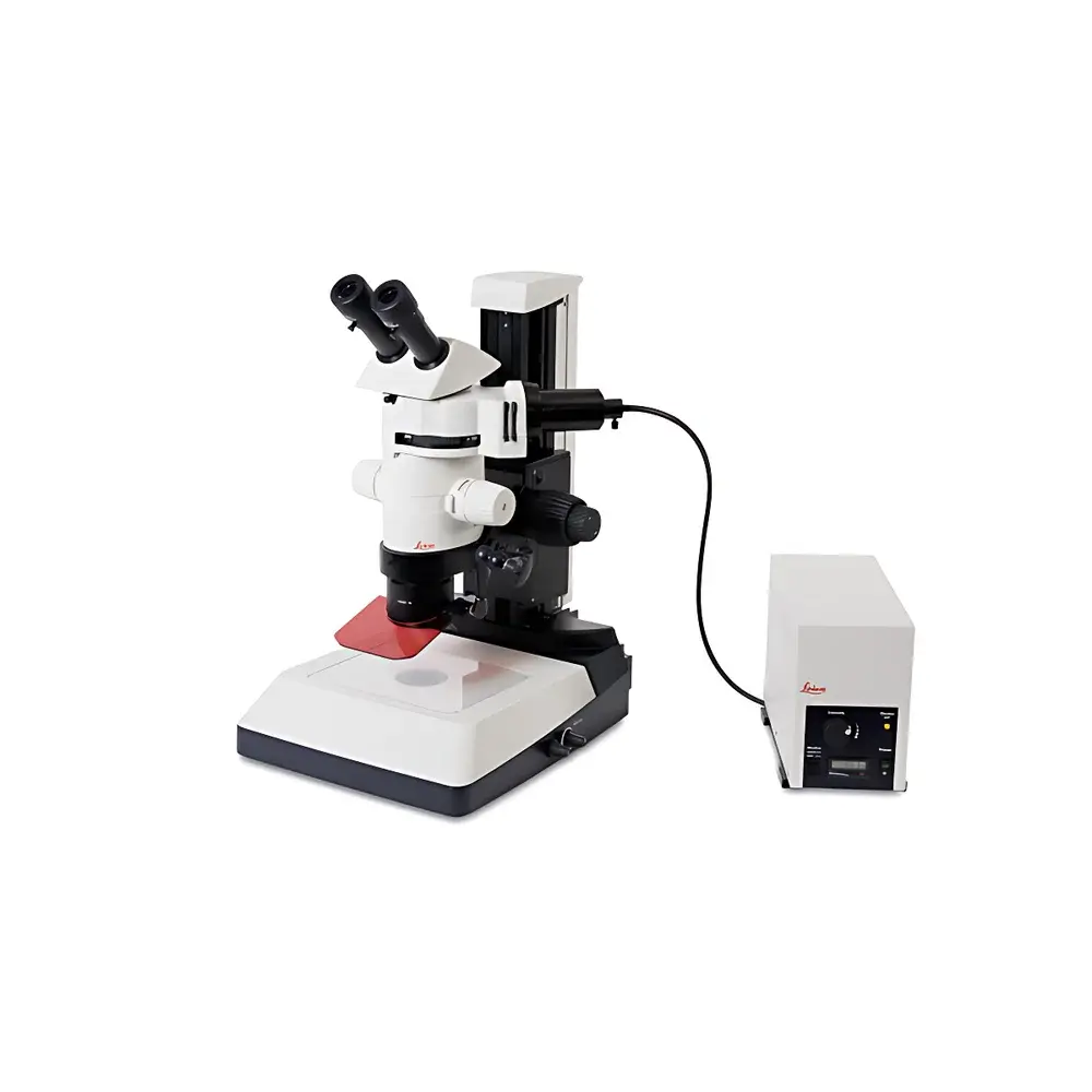
Leica MZ10 F Modular Fluorescence Stereo Microscope
| Brand | Leica |
|---|---|
| Origin | Germany |
| Model | Leica MZ10 F |
| Magnification Range | 8×–80× |
| Resolution | 375 lp/mm |
| Illumination Architecture | Triple-Beam Fluorescence-Specific Optical Path |
| Filter Changer | Leica FluoIII Fast Filter Turret (supports 3 standard or custom filter sets) |
| Compliance | Designed for ISO 10993-compliant sample handling workflows |
Overview
The Leica MZ10 F is a high-performance modular stereo microscope engineered specifically for demanding fluorescence imaging applications in life science research laboratories. Built upon Leica Microsystems’ proven Greenough optical architecture, it integrates a dedicated triple-beam illumination pathway—separating excitation, emission, and transmitted-light paths—to eliminate crosstalk and maximize signal-to-noise ratio in low-light fluorescent specimens. Unlike conventional stereo microscopes that retrofit fluorescence capability, the MZ10 F embeds fluorescence optimization at the optical design level: its beam-splitting optics, chromatic correction, and light-tight housing are all calibrated to preserve quantum efficiency across UV–visible excitation bands (340–650 nm). The system delivers continuous zoom magnification from 8× to 80× with an effective resolution of 375 line pairs per millimeter—validated per ISO 19012-2 standards—enabling subcellular feature discrimination in intact tissues, zebrafish embryos, Drosophila larvae, and organotypic explants.
Key Features
- Triple-beam optical path architecture: Independent excitation, emission, and white-light illumination channels ensure zero spectral bleed-through and optimal contrast in multicolor fluorescence experiments.
- Leica FluoIII fast filter turret: Motorized 3-position filter changer with <150 ms switching time; supports standardized FITC/TRITC/Cy5 cubes or user-defined combinations including DAPI/GFP/mCherry dual- or triple-band configurations.
- Modular platform design: Interchangeable objective modules (e.g., PlanApo 1×, 1.6×, 2.0×), auxiliary magnification changers, and motorized focus drives enable configuration scalability—from routine screening to high-content phenotyping.
- Integrated LED illumination system: Stable, air-cooled high-power LEDs (365 nm, 470 nm, 555 nm, 625 nm) with intensity control (0–100% in 1% increments) and TTL synchronization for time-lapse or triggered acquisition.
- Ergonomic and regulatory-ready hardware: Anti-static coating, ISO 13406-2 compliant viewing optics, and CE/IEC 61000-4-3 certified electromagnetic compatibility for use in shared core facilities and regulated environments.
Sample Compatibility & Compliance
The Leica MZ10 F accommodates a broad range of biological and material science specimens without requiring coverslipping or sectioning—including live zebrafish embryos (up to 72 hpf), mouse brain slices (300 µm thick), plant root tips, polymer scaffolds, and microfluidic devices. Its large working distance (up to 110 mm at 8×) and adjustable stage height support manipulator-integrated workflows (e.g., patch-clamp, microinjection, laser ablation). From a compliance standpoint, the system meets ISO 13485 design controls for medical device R&D and supports FDA 21 CFR Part 11 compliance when paired with Leica LAS X software featuring electronic signatures, audit trails, and role-based access control. All optical components are manufactured in Wetzlar, Germany, under DIN EN ISO 9001:2015 quality management protocols.
Software & Data Management
Leica LAS X Core software provides native acquisition, annotation, measurement (length, area, angle, fluorescence intensity profiling), and export (TIFF, JPEG2000, OME-TIFF) capabilities. For advanced quantification, optional LAS X Advanced Analysis extends functionality to Z-stack projection, multi-channel co-localization (Pearson’s r, Mander’s coefficients), and batch processing via Python API integration. Raw image metadata—including exposure time, gain, filter position, and objective ID—is embedded in EXIF and OME-XML headers, ensuring traceability for GLP audits. Data storage follows FAIR principles: automatic folder structuring by date/user/experiment, configurable backup to network-attached storage (NAS) or LIMS-compatible endpoints.
Applications
- Developmental biology: Real-time tracking of GFP-tagged morphogen gradients in avian embryos or neural crest migration in Xenopus laevis.
- Transgenic model screening: Rapid identification of fluorescent reporter expression patterns in C. elegans, Arabidopsis thaliana seedlings, or CRISPR-edited mouse pups.
- Neuroscience: Stereotactic injection site verification, axon tracing with DiI labeling, and post-hoc correlation with histological sections.
- Quality control in biomanufacturing: Visual inspection of 3D cell aggregates, spheroid uniformity assessment, and viability staining (Calcein AM/Propidium Iodide) in bioreactor harvest samples.
- Materials science: Fluorescent nanoparticle distribution mapping in composite hydrogels or degradation monitoring of labeled polymeric implants.
FAQ
What fluorescence filter sets are included by default with the Leica MZ10 F?
The base configuration includes one FluoIII turret pre-loaded with standard DAPI/FITC/TRITC filter sets. Additional cubes (e.g., Cy5, mCherry, far-red optimized) are available as catalog options.
Is the MZ10 F compatible with third-party cameras?
Yes—via C-mount interface (23.2 mm flange distance) and GenICam-compliant drivers; tested with Hamamatsu ORCA-Fusion BT, PCO.edge 5.5, and FLIR BFS-U3-16S2M-CS models.
Can the system be integrated into automated imaging workflows?
Fully supported through Leica’s SDK (Software Development Kit) and RS-232/Ethernet command protocol for triggering acquisition, filter changes, and focus adjustment from external controllers or LabVIEW environments.
Does the MZ10 F meet requirements for ISO/IEC 17025-accredited testing laboratories?
Yes—when operated with documented calibration certificates (available for objective magnification, illumination uniformity, and camera pixel size), it satisfies clause 6.4.10 on equipment suitability for accredited fluorescence-based morphology assessments.


