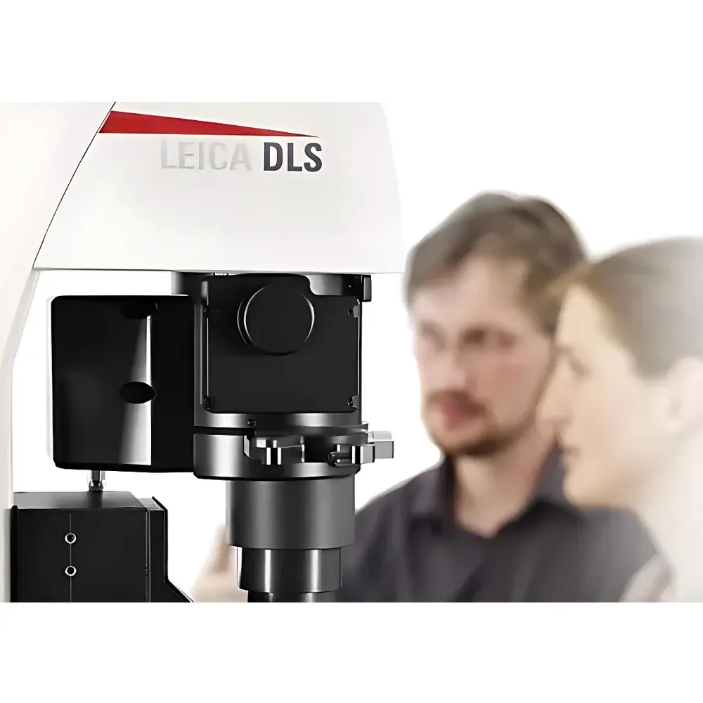
Leica TCS SP8 DLS Digital Light Sheet Microscope
| Brand | Leica |
|---|---|
| Origin | Germany |
| Model | TCS SP8 DLS |
| Imaging Principle | Orthogonal light-sheet illumination integrated with confocal detection |
Overview
The Leica TCS SP8 DLS is a fully integrated digital light sheet microscope engineered to extend the capabilities of the Leica TCS SP8 inverted confocal platform without compromising its core confocal performance. Unlike conventional light sheet systems requiring dedicated optical architectures with orthogonal illumination and detection axes, the TCS SP8 DLS implements a proprietary TwinFlect mirror assembly that deflects the excitation light sheet by 90° into the vertical axis of the existing inverted microscope frame. This enables true orthogonal light-sheet illumination—where the thin, planar excitation beam intersects the detection path perpendicularly—while preserving full access to all confocal scanning modes, spectral unmixing, and resonance scanning functionality. The system operates on the fundamental principle of selective plane illumination microscopy (SPIM): only the focal plane is illuminated, minimizing out-of-focus photon exposure and thereby reducing phototoxicity and photobleaching by up to two orders of magnitude compared to widefield or point-scanning confocal imaging. This makes the TCS SP8 DLS particularly suited for long-term, high-temporal-resolution 3D imaging of live, light-sensitive specimens—including developing embryos, organoids, and neural tissues—under physiologically relevant conditions.
Key Features
- Seamless dual-modality operation: switch between confocal and light sheet modes within the same LAS X software environment without hardware reconfiguration
- TwinFlect mirror technology enabling vertical light-sheet delivery while retaining full confocal optical train integrity
- Dual-sided illumination design eliminates shadowed regions and improves signal uniformity across large fields of view
- Native integration of high-speed sCMOS cameras: pco.edge and Hamamatsu ORCA-Flash 4.0 V2, supporting acquisition rates up to 120 fps at full sensor resolution
- LightSheet Wizard—a guided workflow module in LAS X—for automated light-sheet calibration, thickness optimization, and multi-position acquisition scheduling
- Real-time xytz volumetric acquisition mode for capturing dynamic biological processes in four dimensions (3D + time)
- Modular optical path design accommodating multiple illumination objectives (HC PL Fluotar 2.5×/0.07, HCX PL FLUOTAR 5×/0.15) and detection objectives (HC FLUOTAR L 25×/0.95 W, HC APO L 10×/0.3 W water immersion)
Sample Compatibility & Compliance
The TCS SP8 DLS supports a broad range of live biological samples—from single-cell suspensions and spheroids to intact zebrafish embryos, Drosophila larvae, and mouse brain slices—mounted in standard glass-bottom dishes, agarose-embedded chambers, or custom-designed light-sheet sample holders. Its low-irradiance illumination strategy meets the physiological requirements of developmental biology, neuroimaging, and regenerative medicine studies where sample viability must be maintained over hours or days. From a regulatory standpoint, LAS X software supports user-level access control, electronic signatures, and full audit-trail logging compliant with FDA 21 CFR Part 11 requirements when configured in validated environments. Routine calibration procedures—including light-sheet thickness verification, intensity homogeneity mapping, and Z-stage linearity validation—are documented within the LAS X protocol framework, facilitating adherence to ISO/IEC 17025 and GLP/GMP quality management systems.
Software & Data Management
LAS X serves as the unified control and analysis platform for both confocal and light sheet operations. Its modular architecture includes the LightSheet Wizard for intuitive setup of illumination geometry, multi-angle fusion, and time-lapse scheduling; the 3D Visualization Module for GPU-accelerated rendering, volume clipping, and stereoscopic display; and the Tile Scan & “Mark & Find” modules for automated multi-position imaging across large-area specimens. All acquired datasets are stored in standardized, metadata-rich .lif format, ensuring interoperability with third-party analysis tools including Imaris, Arivis Vision4D, and Fiji/ImageJ. Raw image streams can be exported in TIFF or HDF5 containers with embedded spatial calibration, channel definitions, and acquisition timestamps—critical for reproducible quantitative analysis in peer-reviewed publications and regulatory submissions.
Applications
- Long-term 4D imaging of cardiac dynamics in zebrafish embryos, resolving systolic/diastolic cycles at sub-second temporal resolution
- High-fidelity tracking of cell migration and division during gastrulation and neurulation in Drosophila and Xenopus models
- Volumetric calcium imaging in intact brain slices or cleared tissue preparations using GCaMP or jRGECO reporters
- Multi-region monitoring of organoid maturation and structural remodeling under pharmacological perturbation
- Correlative imaging workflows: initial confocal-guided region-of-interest selection followed by gentle light sheet–based time-lapse acquisition
- Photomanipulation experiments combining optogenetic stimulation (via confocal ROI targeting) with subsequent low-dose light sheet observation
FAQ
Can the TCS SP8 DLS be retrofitted onto an existing Leica TCS SP8 system?
Yes—the DLS module is designed as a field-upgradeable option for qualified TCS SP8 inverted platforms equipped with appropriate scanner and detector configurations.
Does the system support multi-view fusion for improved isotropic resolution?
Yes—LAS X LightSheet Wizard includes online and offline multi-view fusion algorithms that align and merge data from dual-sided illumination paths to correct for absorption artifacts and enhance lateral and axial signal uniformity.
What level of spatial calibration traceability does the system provide?
All objective lenses are factory-calibrated with certified magnification and working distance values; Z-stage linearity is verified using NIST-traceable interferometric standards, and calibration reports are exportable within LAS X.
Is the sCMOS camera interface compatible with third-party acquisition software?
While native control is exclusively through LAS X, raw frame buffers can be accessed programmatically via Leica’s SDK (LSDK) for integration into custom Python- or MATLAB-based analysis pipelines.
How is phototoxicity quantified and minimized during light sheet acquisition?
LAS X provides real-time power monitoring at the sample plane, integrated with exposure time and scan speed controls; users can define maximum permissible fluence thresholds per timepoint, triggering automatic adjustment of laser power or camera gain to stay within biologically safe limits.


