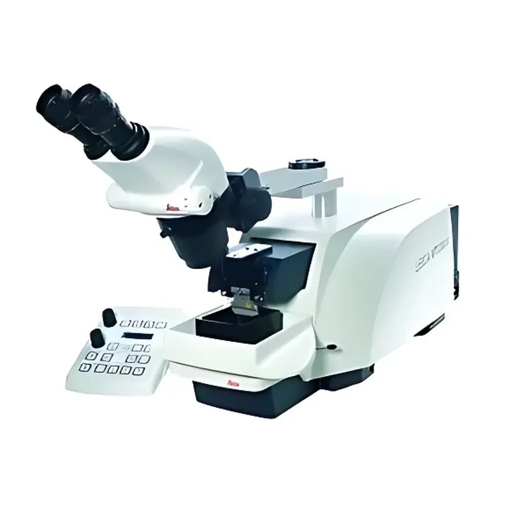
Leica VT1200 S Vibratome
| Brand | Leica |
|---|---|
| Origin | Germany |
| Model | VT1200 S |
| Maximum Sample Size | 33 × 40 mm |
| Section Thickness Range | 0–20,000 µm |
| Sectioning Increment | 1 µm |
| Category | Vibrating Microtome for Fresh & Fixed Tissue |
Overview
The Leica VT1200 S Vibratome is an advanced, high-precision vibrating microtome engineered specifically for the preparation of live-cell-compatible tissue sections in neuroscience and electrophysiology research. Unlike rotary or cryo-microtomes, the VT1200 S operates on the principle of controlled vertical oscillation—applying a precisely regulated sinusoidal vibration to the blade while advancing the specimen at a defined linear speed. This dynamic cutting mechanism minimizes shear stress and compression artifacts, preserving cellular integrity, membrane potential stability, and synaptic ultrastructure in freshly excised or lightly fixed brain, spinal cord, and peripheral nervous system tissues. Developed in close collaboration with Prof. Dr. Peter Jonas’ laboratory at the Institute of Physiology, University of Freiburg, the instrument meets the stringent requirements of patch-clamp recording, calcium imaging, and multi-electrode array (MEA) experiments where surface-layer viability is critical.
Key Features
- Vibrocheck™ Blade Alignment Monitoring System: Real-time optical detection of longitudinal blade deviation during sectioning; integrated feedback enables dynamic correction to maintain sub-micron vertical alignment stability.
- Ultra-Fine Sectioning Control: Adjustable section thickness from 0 µm (zero-cut mode) up to 20,000 µm in 1 µm increments—enabling both ultrathin (e.g., 50–200 µm) live-tissue slices and thick-section histology (e.g., 1–5 mm).
- Dual-Mode Operation: Supports both semi-automatic (user-triggered advance) and fully automatic (programmed sequence) sectioning protocols, configurable via intuitive front-panel interface.
- 8-Profile Memory Function: Stores user-defined parameter sets—including vibration amplitude, frequency, advance speed, section thickness, and pause intervals—for rapid recall and reproducible workflow execution.
- Permanent Magnet Sample Stage: Provides secure, repeatable sample positioning with minimal mechanical disturbance; compatible with agarose-embedded, gelatin-embedded, and PFA-fixed specimens mounted on standard specimen carriers.
- Optimized Fluid Environment Integration: Designed for submerged operation in chilled, oxygenated artificial cerebrospinal fluid (aCSF); includes integrated reservoir ports and temperature-stabilized stage cooling interface.
Sample Compatibility & Compliance
The VT1200 S accommodates specimens up to 33 mm (W) × 40 mm (D), supporting coronal, sagittal, and horizontal brain slice preparations from rodent, primate, and human post-mortem tissue (with appropriate ethical approvals). It is validated for use with paraformaldehyde-fixed, glutaraldehyde-perfused, and unfixed fresh tissue blocks embedded in low-melting-point agarose (2–4 % w/v). The system conforms to IEC 61010-1:2010 safety standards for laboratory electrical equipment and supports GLP-compliant documentation when paired with Leica Application Suite (LAS X) software. While not FDA-cleared as a diagnostic device, its performance aligns with ASTM E2917-21 (Standard Practice for Validation of Analytical Methods in Neuroscience Research) and ISO/IEC 17025:2017 requirements for method validation in academic core facilities.
Software & Data Management
The VT1200 S operates independently via its embedded microcontroller-based interface but integrates seamlessly with Leica LAS X software (v3.7+) for remote configuration, protocol synchronization, and audit-trail generation. All sectioning parameters—including timestamped start/stop events, vibration settings, and thickness logs—are exportable as CSV or XML for traceability. When deployed in regulated environments, optional LAS X modules support 21 CFR Part 11 compliance, including electronic signatures, role-based access control, and immutable audit trails for QC/QA documentation.
Applications
- Preparation of viable acute brain slices for whole-cell patch-clamp electrophysiology.
- Generation of thick-section immunohistochemistry specimens with preserved antigenicity.
- Serial sectioning of hippocampal or cortical explants for multi-photon time-lapse imaging.
- Sectioning of organotypic cultures and tumor spheroids for functional assay integration.
- Correlative light-electron microscopy (CLEM) workflows requiring structural fidelity across modalities.
FAQ
What types of tissue can be sectioned using the VT1200 S?
Fresh, lightly fixed, or cryoprotected neural, cardiac, and epithelial tissues—especially those requiring physiological integrity for functional assays.
Is the VT1200 S compatible with third-party imaging chambers or perfusion systems?
Yes; standardized mounting interfaces and fluid port dimensions allow integration with common perfusion rigs (e.g., Warner Instruments RC-26G, ALA Scientific BSC-200) and upright/dual-path microscope stages.
Does the Vibrocheck™ system require periodic calibration?
No routine recalibration is needed; the system performs self-diagnostic checks at power-on and during idle periods, logging alignment status in the event log.
Can section thickness be adjusted during an ongoing series?
Yes—thickness changes are permitted mid-series without interrupting the vibration cycle, enabling gradient-thickness protocols for layered tissue analysis.
What maintenance is required for long-term reliability?
Bi-weekly cleaning of the blade holder and fluid chamber; annual inspection of the piezoelectric actuator performance by authorized Leica service engineers.


